You can Download Chapter 18 Body Fluids and Circulation Questions and Answers, 1st PUC Biology Question Bank with Answers, Karnataka State Board Solutions help you to revise complete Syllabus and score more marks in your examinations.
Karnataka 1st PUC Biology Question Bank Chapter 18 Body Fluids and Circulation
1st PUC Biology Body Fluids and Circulation NCERT Text Book Questions and Answers
Question 1.
Name the components of the formed elements
In the blood and mention one major function of each of them.
Answer:
Erythrocytes, leucocytes and platelets are collectively called formed elements of the blood. Erythrocytes are the red blood cells (RBC). These molecules play a significant role in transport of respiratory gases. Leucocytes are the white blood cells (WBC). There are two main categories of WBCs granulocytes and agranulocytes. Neutrophils, eosinophils and basophils are different types of granulocytes, while lymphocytes and monocytes are the agranulocytes.
Neutrophils and monocytes are phagocytic cells which destroy foreign organisms entering the body. Basophils secrete histamine, serotonin, heparin etc. and are involved in inflammatory reactions. Lymphocytes are responsible for immune responses of the body. Platelets also called thrombocytes,.are cell fragments produced from megakaryocytes. Platelets are responsible for clotting or coagulation of blood.
Question 2.
What is the Importance of plasma proteins?
Answer:
Fibrinogen, globulins and albumins are the major plasma proteins. Fibrinogen are needed for clotting or coagulation of blood. Globulins primarily are involved in defense mechanisms of the body. Albumins help in osmotic balance.
Question 3.
Match Column I with Column II:
Column I – Column II
(a) Eosinophils – (i) Coagulation
(b) RBC – (ii) Universal Recipient
(c) AB Group – (iii) Resist Infections
(d) Platelets – (iv) Contraction of Heart
(e) Systole – (v) Gas transport
Answer:
(a) – (iii)
(b) – (v)
(c) – (ii)
(d) – (i)
(e) – (iv)
Question 4.
Why do we consider blood as a connective tissue?
Answer:
Connective tissue usually are involved in structure and support and derived from mesoderm. Blood is considered as a connective tissue because:
- It has the same origin (mesodermal) as do the other connective tissues.
- blood connects the body systems together bringing the needed oxygen, nutrients, hormones and other important molecules and removing the waste.
Question 5.
What is the difference between lymph and blood?
Answer:
| Blood | Lymph |
| (i) Red in colour (ii) Contains RBC (iii) High protein concentration (iv) Transportation of gases, nutrients, and waste | (i) Colourless liquid (ii) Does not contain RBC (iii) Low protein concentration (iv) Contains specialised lymphocytes which are responsible for defense of the body. |
Question 6.
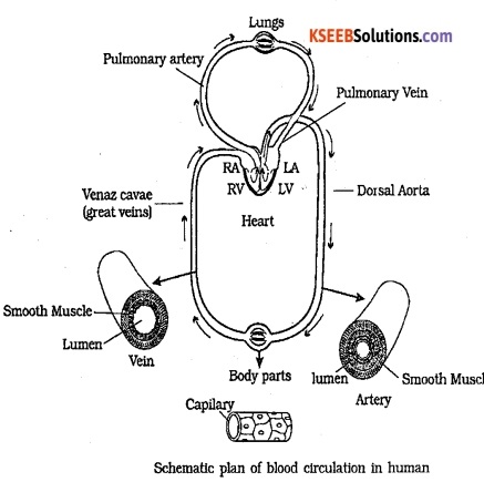
What is meant by double circulation? What is its significance?
Answer:
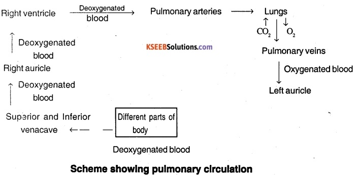
The double circulation refers to the separate systems of pulmonary circulation and systemic circulation. The deoxygenated blood pumped into the pulmonary artery is passed on to the lungs from where the oxygenated blood is carried by the pulmonary veins into the left atrium. This pathway constitutes the pulmonary circulation.
The oxygenated blood entering the aorta is carried by arteries and capillaries to the tissues from where the deoxygenated blood is collected by a system of venules, veins and vena cava and emptied to the right atrium. This is the systemic circulation.
The systemic circulation is responsible for providing nutrients, O2 and other essential substances to the tissues and taking away CO2 and other harmful substances for elimination.
![]()
Question 7.
Write the differences between:
(a) Blood and Lymph
(b) Open and Closed system of circulation
(c) Systole and Diastole
(d) P-wave and T-wave
Answer:
(a) Blood is a fluid connective tissue that contains plasma, RBC, WBC and platelets. Lymph is a tissue fluid formed from blood. It contains only lymphocytes.
(b) Open circulatory system is present in arthropods and molluscs in which blood pumped by the heart passes through large vessels into open spaces or body cavities called sinuses.
Closed circulatory system is present in annelids and chordates in which blood pumped by the heart is always circulated through a closed network of blood vessels. Closed system can be better regulated than open system.
(c) Systole is the contraction of heart muscle and diastole is the dilation of the heart muscle. Systole results in increased pressure in heart chambers, whereas diastole results in decreased pressure.
(d) The P – wave represents the electrical excitation (on depolarisation) of the atria, which leads to the contraction of both the arteria. The T – wave represents the return of the ventricles from excited state to normal state (repolarisation). The end of T – wave marks the end of systole.
Question 8.
Describe the evolutionary change in the pattern of heart among the vertebrates.
Answer:
All vertebrates possess a muscular chambered heart. Fishes have 2 – chambered heart with an atrium and a ventricle. They have single circulation in which heart pumps deoxygenated blood, which is oxygenated by gills and supplied to body parts. Amphibians and reptiles have a 3 – chambered heart with two atria and a single ventricle.
They possess incomplete double circulation with oxygenated and deoxygenated blood getting mixed up in a single ventricle. Crocodiles, birds and mammals possess a 4 chambered heart with two atria and two ventricles. Two separate circulatory pathways are present in these organisms and hence have double circulation.
Question 9.
Why do we call our heart myogenic?
Answer:
Normal activities of the heart are regulated intrinsically i.e., auto regulated by specialised muscles, hence our heart is called myogenic.
Question 10.
Sino-atrial node is called the pacemaker of our heart. Why?
Answer:
In human heart, Sino atrial node initiates the conduction of heart beat. The excitory wave of SA node is called cardiac impulse and it determines the rate of heart beat and sets the pace of the activities of the heart. Hence Sino atrial node is termed as pacemaker of the heart.
Question 11.
What is the significance of atrio ventricular node and atrio-ventricular bundle in the functioning of heart?
Answer:
The heart beat initiated by Sino atrial node is picked up by the Atrio – ventricular node and the action potential is conducted to the ventricular side. From the AV node the heart beat is transmitted to the AV bundle (Bundle of HiS) which transmits it through the entire ventricular musculature.
Question 12.
Define a cardiac cycle and the cardiac output.
Answer:
The sequential events that occur from the beginning of one heart beat to the beginning of the next heart beat which is cyclically repeated is called the cardiac cycle. The volume of blood pumped by the ventricle per minute is called the cardiac output. It ranges about 5000 ml (5 L) in a healthy individual.
Question 13.
Explain heart sounds.
Answer:
During each cardiac cycle two prominent sounds lub and dub are heard. The first heart sound (lub) is associated with the closure of the tricuspid and bicuspid valves which extends for 0.16 – 0.9 sec. The second heart sound (dub) is associated with the closure of the semilunar valves and extends for 0.10 sec.
Question 14.
Draw a standard ECG and explain the different segments in it.
Answer:
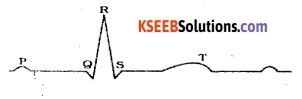
Diagrammatic presentation of a standard ECG
Each peak in the ECG is identified with a letter from P to T that corresponds to a specific electrical activity of the heart. The P – wave represents the electrical excitation (or depolarisation) of the atria which leads to contraction of both the atria.
The QRS complex represents the depolarisation of the ventricles, which initiates the ventricular contraction. The contraction starts shortly after Q and marks the beginning of the systole.
The T – wave represents the return of the ventricles from excited to normal state (repolarisation). The end of the T – wave marks the end of systole.
1st PUC Biology Body Fluids and Circulation Additional Questions and Answers
1st PUC Biology Body Fluids and Circulation One Mark Questions
Question 1.
What is the Coagulation of Blood?
Answer:
When blood has been shed, it quickly becomes sticky and soon sets as a red jelly. This jelly or clot contracts or shrinks, and a straw coloured fluid called serum is squeezed out from it.
Question 2.
What is fibrinogen?
Answer:
It is a soluble plasma protein which is acted upon by thrombin to form insoluble fibrin clot.
Question 3.
What is Prothrombin?
Answer:
This is an inactive precursor of thrombin formed in the liver.
Question 4.
What is the function of factor VI?
Answer:
It is required for the formation of prothrombin activator by tissue extract.
Question 6.
Name the instrument used for measuring blood pressure. (Apr. 1983, Sep. 91, July. 06)
Answer:
Sphygmomanometer.
Question 7.
Expand S.A.N. (April 86)
Answer:
Sino-Atrial Node.
Question 8.
What is pulse? (April 86)
Answer:
The pressure waves created by the heart beat along the arteries when the left ventricle pumps nearly 70 ml. of blood each systole into the aorta is called as the pulse.
![]()
Question 9.
What is blood pressure? (M.Q.P., Oct. 86, 94)
Answer:
Blood pressure is the force with which blood pushes against the wall of the blood vessels (arteries) and is generated by the cardiac output.
Question 10.
Where is tricuspid valve located? (April 87)
Answer:
The tricuspid valve is located between the right Atrium (auricle) and right ventricle.
Question 11.
How much is the diastolic pressure of a normal adult human being? (Oct. 87)
Answer:
It is about 80 mm of Hg (mercury)
Question 12.
State the normal blood pressure of man.
Answer:
‘120/80’ (April 89, 91)
Question 13.
What type of heart is found in Man? (April 90, Oct. 93)
Answer:
The heart of man is of the ‘Myogenic type’.
Question 14.
Name the blood vessels from which the heart receives oxygenated blood?
Answer:
Pulmonary Veins. (April 92, 95)
Question 15.
Which is the valve present in between the left auricle and left ventricle? (April 97, M.Q.P.)
Answer:
The Bicuspid or mitral valve.
Question 16.
Mention the meaning and causes of the Myocardial infraction (M.Q.P.)
Answer:
‘Myocardial infraction means death of cardiac muscle in certain locations of the heart due to faulty coronary supply of blood or circulation. It is commonly called ‘Heart Attack’.
It may be caused due to thrombus (or blood clots of different forms) in one of the coronary arteries cutting off blood supply to certain region of the heart (myocardium). Thrombosis may be caused due to hypertension, high cholesterol, levels, smoking & Diabetes mellitus.
![]()
Question 17.
Where does the impulse for the heart beat originate? (Oct. 89)
Answer:
The pacemaker or Sinu-Atrial node (SAN)
Question 18.
Expand SAN and ET. (April 98)
Answer:
- San – Sino Atrial Node (Pace Maker)
- Et – Embryo Transfer.
Question 19.
What is myocardial infarction? (April 00,06)
Answer:
Myocardial infarction/ heart attack is the death of cardiac muscle due to faulty coronary blood supply or circulation.
Question 20.
Name the valve present in between aorta and left ventricle. (Oct. 2000)
Answer:
Bicuspid valve or mitral valve.
Question 21.
What is pericardium? (Oct. 2002)
Answer:
The pericardium is the tough connective tissue sac enclosing the heart and holding it in place.
Question 22.
Name the artery which carries deoxygenated blood. (Oct. 2002)
Answer:
pulmonary trunk.
Question 23.
What are coronary arteries? (Oct. 2003)
Answer:
Coronary arteries are the arteries arising from the aorta and supply blood to the myocardium of the heart.
Question 24.
Give an equation for cardiac output. (M.Q.P., July 2006)
Answer:
Cardiac output is the product of stroke volume and heart rate/minute.
Equation for cardiac out put is,
CO = SV x HR i.e = 70 x 72 = 5040ml.
CO is cardiac output, SV = Stroke Volume HR = heart rate (Number of heart beats/minute).
Question 25.
Write the reason for Cyanosis. (April 2007)
Answer:
Tetralogy of fallot (interventricular septal defect).
Question 26.
Name the enzyme which converts fibrinogen into fibrin. (March 2008)
Answer:
Thrombin.
Question 27.
Define cardiac output. (July 2008)
Answer:
Cardiac output is the volume of blood ejected out from the ventricles over one minute.
Question 28.
Define stroke volume. (March 2009)
Answer:
Stroke volume is the amount of blood ejected by the ventricle per heartbeat.
Question 29.
Give reason:
Human heart is myogenic. (March 2009)
Answer:
Human heart is myogenic because the heart beat originates in the heart itself.
Question 30.
What is double circulation? Mention its types. (March 2011)
Answer:
The circulation of blood in which blood passes through the heart twice during one complete circuit is called double circulation. The types are pulmonary and systemic circulation.
Question 31.
Ventricles are thicker than atria. (March 2011)
Answer:
The two ventricles represent the pumping chambers of the heart, hence are thicker than auricles.
Question 32.
Name the type of granulocytes that play an important role in inflammatory reactions.
Answer:
Basophils
![]()
Question 33.
Name the type of granulocytes that are significant In allergic reactions and detoxification.
Answer:
Eosinophils.
Question 34.
What transmits the cardiac impulse from the atria to the ventricles? (Delhi 2000 C)
Answer:
Atrio – ventricular bundle.
Question 35.
Which of the four chambers of the human heart has the thickest muscular walls?
Answer:
Left ventricle.
Question 36.
Name one animal in which heart pumps only deoxygenated blood. How many chambers does this heart have? (All India 1998 C)
Answer:
Fish; the heart has two chambers.
Question 37.
What is joint diastole?
Answer:
Joint diastole is the phase in the cardiac cycle during which both atria and ventricles are relaxed simultaneously.
Question 38.
What is the function of sino – atrial node?
Answer:
Sino – atrial node generates the action potential and determines the rate of the heart.
Question 39.
What causes the first heart sound? (Foreign 1997)
Answer:
The closure of AV – valves.
Question 40.
Name the two fluids circulated in the body of mammals
Answer:
Blood and lymph
Question 41.
What is blood?
Answer:
Blood is a connective tissue consisting of ‘ a fluid matrix, plasma and formed elements.
Question 42.
Where are RBCs formed from In an adult human?
Answer:
RBCs are formed from the red bone marrow.
Question 43.
Why are erythrocytes red in colour?
Answer:
Erythrocytes contain haemoglobin which gives it a red colour.
Question 44
How many RBCs are present in a mm3 of blood of an adult human?
Answer:
5.0 – 5.5 millions.
Question 45.
Name the leucocytes that are phagocytic.
Answer:
Neutrophils and monocytes.
Question 46.
Name any two substances secreted by basophils.
Answer:
Histamine, serotonin, heparin etc.
Question 47
Name the cells which produce thrombocytes.
Answer:
Mega karyocytes of bone marrow.
Question 48.
Why is blood group ‘O’ called a universal donor?
Answer:
Blood group ‘O’ has no antigen to react with the antibodies of the recipient. So it can be transfused to any person and is called as universal donor.
Question 49.
Why is blood group AB called as universal recipient?
Answer:
Blood group AB does not have any anti-body to react with the antigen of donor, and can accept any blood group. So it is known as universal recipient.
Question 50.
Name the element Involved in blood clotting.
Answer:
Calcium.
Question 51.
Name a reptile that has four chambered heart.
Answer:
Crocodile.
Question 52.
Name the double layered membranous covering of the heart.
Answer:
Pericardium.
![]()
Question 53.
Where is pericardial fluid present?
Answer:
Pericardial fluid is present between the two layers of the pericardium.
Question 54.
What Is the average number of heart beats in a man?
Answer:
10 – 75 per minute. (72 is average)
Question 55.
Name the phase of the cardiac cycle in which both atrlo and the ventricles are relaxed simultaneously.
Answer:
Join diastole.
Question 56.
What are Purkinje fibers?
Answer:
The minute fibres throughout the ventricular musculature that arise from the branches of the right and left atrioventricular (AV) bundles on respective sides are called as’ Purkinje fibres.
Question 57.
What is the duration of one cardiac cycle?
Answer:
About 0.8 seconds.
Question 58.
Name two organs affected by high BP.
Answer:
Brain and Kidney.
Question 59.
What is the main symptom of heart failure?
Answer:
Congestion of lungs.
Question 60.
What is the organ heart derived from?
Answer:
Mesoderm.
Question 61.
Why nodal musculature is called auto excitable?
Answer:
The nodal musculature has the ability to generate action potentials without any external stimuli.
Question 62.
What is lymph?
Answer:
Lymph is a colourless liquid present in the lymphatic system containing specialised lymphocytes which are responsible for immune system of the body.
Question 63.
Name two classes with open circulatory system.
Answer:
Arthropods and molluses.
Question 64.
Who are called Rh positive and Rh negative?
Answer:
Individuals having Rh antigen on the surface of their RBCs are called Rh positive and the one who do not contain these antigens are called Rh-negative.
Question 65.
What separates the atrium and the ventricle of the same side?
Answer:
Atrio – ventricular septum.
1st PUC Biology Body Fluids and Circulation Two Marks Questions
Question 1.
Define Pacemaker. Give an example. (April 93)
Answer:
The pacemaker is a compact mass of muscle fibers which initiates each cardiac cycle, thereby setting or establishing the rate of beating-(pace) of the heart.
Eg: The sinoatrial (Sinuatrial) node situated inferior to the opening of the superior vena cava.
Question 2.
Define Hypotension and Hypertension. (Oct. 93)
Answer:
- Hypotension or Low blood pressure is a clinical condition wherein the SBP (Systolic Blood Pressure) and DBP (Diastolic Blood Pressure) fall below the normal value, i.e below 100 and 50 mmHg respectively.
- Hypertension or High blood pressure is a clinical condition wherein the DBP rises or elevates above the normal value (i.e. more than 90 mm Hg).
Question 3.
Explain pulmonary circulation. (April 97, 2000, 2007)
Answer:
In the pulmonary circulation (or route) the blood leaves the right ventricle of the heart through the pulmonary artery (trunk), carrying deoxygenated blood to the lungs through its left and right branches. After oxygenation of this blood in the lungs it is carried back into the left atrium or auricle through the left and right pulmonary veins.
Question 4.
Why is the human circulation called complete double circulation. (M.Q.P.)
Answer:
In the human heart the chambers and blood vessels (arteries; veins) comprising the circulatory system are arranged and organised to form the pulmonary and systemic circulations to receive, pump, purify (or oxygenate) and repump blood from the body cells in such way that they not only keep the oxygenated and deoxygenated blood separately in the heart at a time but also help to circulate blood through the heart twice during one complete circuit, ensuring continuous supply of purified or oxygenated blood to the body. This type of circulation is known as a complete double circulation.
Question 5.
Explain briefly the systemic circulation. (April 2003)
Answer:
The left atrium is the receiving chamber for the oxygenated blood brought to it by the four pulmonary veins (two from each lung). The blood leaves the left atrium to fill the left ventricle which pumps the blood into the aorta.
The aorta is the main artery of the systemic circulation. From it, blood is contributed to the other arteries, arterioles and capillaries to different organs of the body. Blood is collected from the organs and poured into the venae cavae which bring blood back to the right atrium. This route constitutes the systemic circulation.
![]()
Question 6.
What is cardiac output?
Answer:
It is volume of blood ejected out from the heart (ventricles) over one minute. Cardiac output is the product of the stroke volume and the heart rate/ minute.
CO = SV × HR 70 × 72 = 5040 ml.
The cardiac output is influenced by several factors like heart rate, contractility of the heart etc.
Question 7.
What is cardiac cycle? What is heart rate?
Ans:
The alternate systole and diastole of auricles and ventricles constitutes heart beat and cardiac cycle. The number of heart beats / minute is called heart rate.
Question 8.
Mention the artery that carries deoxygenated blood and vein that carries oxygenated blood.
Answer:
Pulmonary artery Pulmonary vein.
Question 9.
Give a schematic representation of Pulmonary circulation.
Answer:
Question 10.
Can all the four chambers of the human heart experience systole simultaneously? Justify your answer.
Answer:
No, all chambers cannot experience systole simultaneously.
Arteriole systole occurs first as it is directly connected to SA-node which initiates the cardiac impulse. This impulse is then discharged to atria and transmitted from atrial muscles to the ventricular muscles through atrio – ventricular node and Av – bundle. The impulse passes slowly across the AV-node and hence ventricular systole begins after the atrial systole is completed.
Question 11.
Name the different types of granulocytes. Give the function of the one which constitutes maximum percentage of the total leucocytes. (Delhi 2004)
Answer:
- Neutrophils, eosinophils and basophils are three types of granulocytes.
- Neutrophils constitute 60 – 65 % of the total leucocytes and are phagocytic cells which destroy foreign organisms entering the body.
Question 12.
Write a note on hepatic portal system.
Answer:
Hepatic portal system is a unique vascular connection that exists between the digestive tract and
the liver.
The hepatic portal vein carries blood from intestine to the liver before it is delivered to the systemic circulation.
Question 13.
Name In a sequence the specialised cardiac muscle fibres responsible for conduction of heart beat. Also mention their location in the heart. (All India 1999 C)
Answer:
Heart conducting system contains Sino-atrial node, Atrio-ventricular node, AV-bundle and Purkinje fibres.
- SA node is situated in the upper lateral wall of the right atrium.
- AV node is located in the posterior part of the interatrial septum.
- The AV bundle branches into two and enter left and right ventricles.
- Purkinje fibres are located throughout the ventricular wall.
Question 14.
Name the events of one complete heartbeat in proper sequence.
Answer:
Joint diastole → atrial systole → ventricular systole and atrial diastole → ventricular diastole.
Question 15.
Differentiate between RBCs and WBCs.
Answer:
| RBC | WBC |
| (a) They do not have nucleus at maturity. (b) They possess haemoglobin and hence red. (c) They help in transport of respiratory gases. (d) They are about 5 million / mm3 of blood. | (a) They contain a large characteristic nucleus. (b) They are colourless and contain no pigment. (c) They help in defence mechanism. (d) They are about 7000 / mm3 of blood. |
Question 16.
Differentiate between open and closed system of circulation
Answer:
| Open circulating system | Closed circulating system |
| (a) Blood flows through open spaces and channels. | (a) Blood flows through heart and closed blood vessels. |
| (b) Sufficiently high blood pressure cannot be maintained | (b) Sufficiently high blood pressure can be maintained |
| (c) The volume of blood flowing to different tissues cannot be regulated. | (c) The volume of blood can be regulated. |
| (d) Blood flows at a slow velocity. | (d) Blood flows at a higher velocity. |
| (e) Body tissues are in direct contact with blood. | (e) Body tissues do not come in contact with blood. |
Question 17.
Where are bicuspid and tricuspid valves located in human? What is their function?
Answer:
- Bicuspid valve guards the opening in the atrio – ventricular septum between the left atrium and left ventricle.
- Tricuspid valve guards the opening in the atrio – ventricular septum between the right atrium and right ventricle. They allow the flow of blood in one direction.
Question 18.
Define stroke volume. What is its value?
Answer:
The volume of blood pumped by a each ventricle during one cycle is called stroke volume. It is about 70 ml.
Question 19.
Name the four types of blood groups. What is the basis for such grouping?
Answer:
The four types of blood groups are A, B, AB and O. Blood grouping is based on the presence or absence of two surface antigens on the RBCs namely A and B.
1st PUC Biology Body Fluids and Circulation Three Marks Questions
Question 1.
Draw a schematic representing double circulation in human.
Answer:
Question 2.
Where and from which cells do platelets originate? What Is their life span? How do they act when blood vessels get injured?
Answer:
Platelets originate from the megakaryocytes in the bone marrow. They live for about seven days. They release thromboplastins, which help convert prothrombin of the plasma into thrombin and thus help in clotting of blood.
Question 3.
Explain the chemical events taking place during clotting /coagulation of blood.
(Foreign 1997)
Answer:
An injury or a trauma stimulates the plate- 195 lets in the blood to release certain factors which activate the mechanism of coagulation. Clot or coagulam informed mainly of a network threads called fibrins in which dead and damaged formed elements of blood are trapped. Fibrins are formed by the conversion of inactive fibrinogens in the plasma by the enzyme thrombin.
Thrombins, in turn are formed from another inactive substance present in the plasma called prothrombin. An enzyme complex, thrombokinase, is required for the above reaction which is formed by a series of linked enzymic reactions involving a number of factors present in the plasma in an inactive state. Calcium ions play a very important role in clotting.
![]()
Question 4.
Mention the functions of each of the following :
(a) Basophils
(b) Eosinophils
(c) Monocytes
Answer:
(a)Basophils: They secrete histamine, serotonin, heparin etc. and are involved in inflammatory reactions.
(b) Eosinophils: They resist infections and are also associated with allergic reactions.
(c) Monocytes: They are phagocytic which destray foreign organisms entering the body.
Question 5.
Why Is it necessary to check the Rh-factor of the blood of a pregnant woman?
Answer:
A Rh – negative person, if exposed to Rh +ve blood, will form specific antibodies against the Rh antigens. A special case of Rh incompatibility has been observed between the Rh -ve blood of a pregeant mother with Rh +ve blood of the foetus. Rh antigens of the foetus do not get exposed to the Rh -ve blood of the mother in the first pregnancy as the two bloods are well separated by the placenta.
During the delivery of the first child, there is a possibility of exposure of the maternal blood to small amounts of the Rh +ve blood from the foetus. In such cases, the mother starts preparing antibodies against Rh antigen in her blood. In case of her subsequent pregnancies, the Rh antibodies from the mother (Rh -ve) can leak into the blood of the foetus (Rh +ve) and destroy the foetal RBCs.
This could be fatal to the foetus or could cause severe anaemia and jaundice to the baby. This condition is called as erythroblastosis foetulis. Hence it is necessary to check the Rh factor of the blood of pregnant woman.
Question 6.
How is cardiac activity regulated?
Answer:
Normal activities of the heart are regulated intrinsically by specialised muscles, and hence the heart is called myogenic. A special neural centre in the medulla oblongata can moderate the cardiac function through Autonomic Nervous System. (ANS).
Neural signals through the sympathetic nerves can increase the rate of heart beat, the strength of ventricular contraction and thereby the cardiac output. Para sympathetic neural signals decrease the rate of heart beat, speed of conduction of action potential and thereby the cardiac output. Adrenal medullary harmones can also increase the cardiac out-put.
Question 7.
Explain any three disorders of circulatory system.
Answer:
(i) High Blood Pressure (Hypertension):
Hypertension is the term for blood pressure that is higher than normal (120/80). High blood pressure leads to heart diseases and also affects vital organs like brain and kidney.
(ii) Coronary Artery Disease (CAD):
Coronary Artery Disease, often referred to as atherosclerosis, affects the vessels that supply blood to the heart muscle. It is caused by deposits of calcium, fat, cholesterol and fibrous tissues, which makes the lumen of arteries narrower.
(iii) Heart Failure:
Heart failure means the state of heart when it is not pumping blood effectively enough to meet the needs of the body. The main symptoms are congestion of the lungs.
1st PUC Biology Body Fluids and Circulation Five Marks Questions
Question 1.
Explain the mechanism of Blood clotting. (Mar. 09)
Answer:
Calcium salts, cell injury which liberates thrombokinase, (thromboplastin) thrombin and fibrin.
Prothrombin + Ca + thrombokinase →
Thrombin.
Thrombin + fibrinogen → Fibrin
Fibrin + bloodcells → Clot (Fibrin clot)
The clotting consists 3 phases.
Stage I: Thromboplastin or thrombokinase is liberated from injured tissue or shed blood.
Stage II: The thromboplastin in the presence of ionic calcium acts on prothrombin, a protein present in the blood plasma and coverts it to thrombin.
Stage III: The thrombin in turn acts on fibrinogen, the soluble protein present in plasma and converts it to fibrin. It is insoluble and precipitates as a net work of elongated thread like fibres enmeshing cellular and liquid components in the mesh work and giving it a solid appearance. Subsequently the fibres shrink an the mesh becomes closely knit. It holds the cellular elements, but exuding out the liquid. The liquid is the serum.
Question 2.
What is conducting system? Explain its role in functioning of the heart.
(Oct. 83, 96, April 90, 94)
Answer:
Blood is pumped by the heart by rhythmic contractions and relaxation (heart beat) of the auricles and ventricles in a definite order.
The conducting system brings about the rhythmic and automatic working of the heart (i.e. contraction and relaxation of the heart) through the transmission of impulses generated within specialised cardiac muscles and its channels, thereby accomplishing continuous circulation (or pumping) of blood through the heart.
The impulses for initiation of heart beat starts in the SAN (Sino-atrial node) or Pacemaker of the heart and is transmitted to the AV node (Atrioventricular node) through 3 special tracts or pathways (composed of specialised cardiac muscle fibres) called internodal pathways. From here the stimulus is carried by a single tract of specialised muscle called the “Bundle of His” in the beginning and diverted to the left and right side of the heart by its branches, (i.e. left and right branches).
These branches later rebranch profusely into Purkinje fibres. The Purkinje’s fibres transmit the Impulses received from the branches of Bundle of His to all the parts of the Ventricles bringing about simultaneous contraction of Ventricle pumping blood from right ventricle into lungs through pulmonary artery and pumping blood from left ventricle into aorta to various parts of the body through its branches. In this way impulses are transmitted through the conducting system ensuring systematic working of heart and blood supply.
![]()
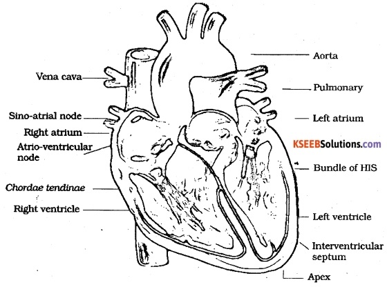
Question 3.
Explain the structure of mammalian heart with a neat labelled diagram.
(Oct. 83, 86, 91,96, 97, April 85, 06, 07)
Answer:
The heart of Mammals is somewhat fist shaped, broader at the anterior end and triangular, narrow & pointed at the posterior end marked by an apex at its tip.
The heart is made up of cardiac muscle and protected on its outer side with a double membrane, the pericardium, with its pericardial fluid. Internally the heart is 4 chambered with a left
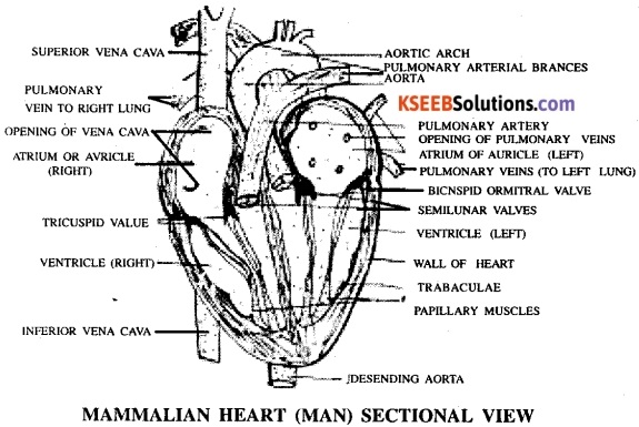
half consisting of the left auricle (receiving chamber) followed by the left ventricle (pumping chamber) and the right half consists of the right auricle (receiving chamber) and the right ventricle (pumping chamber). The left half is separated completely from the right half with the auriculoven-tricular septum.
The auricles or atrium (the receiving chambers) of the heart are anterior in position followed by the ventricles which posterior in position. The walls of the auricles are thinner than the ventricles and the walls of the ventricles bear special papillary muscles and tribaculae holding the valves present between the auricles and ventricles. The chambers are lined by simple squamous epithelium or endocardium.
The auricles and ventricles are separated and guarded by valves allowing the flow of blood only in one direction due to their mode of arrangement.
The right auriculo-ventricular value is a tricuspid (3 flapped) valve opening towards the right ventricle and that between the left auricle and ventricle is the Bicuspid (2 flapped) or mitral valve opening towards the left ventricle.
The left side of the heart receives oxygenated blood through a set of blood vessels and pumps blood through a set of blood vessels towards the body. The right side of the heart receives impure (deoxygenated) blood from the body (cells) and pumps blood to lungs for purification through another set of blood vessels.
They are as follows;
(i) Superior and inferior vena cava:
Bring blood collected (deoxygenated) from various (upper NS and lower) parts of the body into the right
auricle.
(ii) Pulmonary artery and its branches: The pulmonary artery carries deoxygenated blood pumped by the right ventricle to the lungs for purification of blood in terms of respiratory gases (removal CO2 & addition of O2) channelised to left & right lobes of the lung with the help of its branches. The pulmonary artery is guarded by semilunar valves.
(iii) The pulmonary veins: Collect the purified blood from the left & right lobes of the lung and carry it into the left auricle.
(iv) Aorta: It is the largest vessel of the heart guarded by semilunar valves.
It transports blood (oxygenated) pumped by the left ventricle (received from left auricle) into the body through its branches (to the upper and lower parts of the body).
![]()
Question 4.
What is double circulation? Describe with reference to a human heart. (April 87, 91, 99, Oct. 95, July 07)
Answer:
The circulation of blood in which blood passes through the heart twice during one complete circuit is called as double circulation.
The heart of man is four-chambered. The left side of the heart is made up of left atrium or auricle (receiving chamber anterior region) and the left ventricle (pumping chamber posterior region). These two chambers are guarded by a bicuspid valve, opening towards the ventricle. The right half of the heart is made up of right atrium or auricle (on the anterior region) and right ventricle (on posterior region) these two chambers are guarded by the tricuspid valves opening towards ventricle. The valves ensure unidirectional flow of blood and prevent its backflow. The right half of the heart receives deoxygenated blood from body
and the left half receives oxygenated blood from the lungs. The two halves are separated by the atrioventricular septum ensuring absolutely no mixing up of the blood between these two halves.
The oxygenated blood leaves the left ventricle through the aorta (the great vessel) and reaches various parts of the body through its branches, arteries and capillaries. After this blood is deoxygenated in cells and takes up CO2, it is returned back into the right side of the heart by venules and veins forming larger veins, the superior and inferior vena cava into the right auricle.
From here it enters the right ventricle and is pumped into the lungs through the pulmonary artery. (Aorta & pulmonary artery are guarded by semilunar valves). Later the oxygenated blood from lungs is, carried back into the left auricle or atrium by pulmonary- veins. From the left auricle it enters the left ventricle and from here blood is pumped into the aorta for distribution.
Thus blood circulates (without mixing up) continuously in the human heart and passes it twice during one complete circuit keeping the impure or deoxygenated blood separate from the oxygenated or pure blood during the circulation. This is known as double circulation.
![]()
Question 5.
Fill in the blanks:
(a) ……………….. is a plasma protein needed for coagulation of blood.
(b) Granulocyte which is phagocytic ………………..
(c) ………………..is the pacemaker of human heart.
(d) Expand ECG: ………………..
(e) Sound associated with closure of the tricuspid and bicuspid valves is ………………..
Answer:
(a) Fibrinogen
(b) Neutrophils
(c) Sino – atrial node (SAN)
(d) Electrocardiograph
(e) lub.
Question 6.
Draw a neat labelled structure (section) of a human heart.
Answer:
Question 7.
Match the Following
| I | II |
| (a) Depolarisation of atria (b) Repolarisation (c) Sympathetic nerve (d) Parasympathetic nerve (e) Depolarisation of ventricles. | (i) Increase cardiac output (ii) Decrease cardiac output (iii) P – wave (iv) QRS complex (v) T -wave. |
Answer:
(a) → (iii)
(b) → (v)
(c) → (i)
(d) →(ii)
(e) → (iv)
Question 8.
Briefly explain cardiac cycle.
Answer:
The sequential event in the heart which is cyclically repeated is called the cardiac cycle and it consists of systole and diastole of both the atria and ventricles. Initially all the four chambers of heart are in a relaxed state, i.e., joint diastole. Tricuspid and bicuspid valves are open resulting in inflow of blood into the left and right ventricles from pulmonary veins and vena cava. Semilunar valves are closed at this stage. SAN (sino-atrial node) generates an action potential resulting in simultaneous contraction of both atria – the atrial systole.
The action potential is conducted to the ventricular side by the AVN and AV bundle from where the bundle of His transmits it through the entire ventricular musculature. This causes contraction of ventricles (ventricular systole) and relaxation of atria (diastole). Ventricular Systole increases the ventricular pressure causing closure of tricuspid and bicuspid valves. As pressure increases, the semilunar valves of pulmonary artery and aorta are forced open, allowing blood to flow through them into circulatory pathways.
The ventricles now relax (ventricular diastole) and semilunar valves close. As the ventricular pressure declines further, tricuspid and bicuspid valves are pushed open by the pressure in the atria. The blood now once again moves freely to the ventricles. The ventricles and atria are now again in a relaxed (joint diastole) state, as earlier and the process continue.
The duration of a cardiac cycle is 0.8 seconds and during this period each ventricle pumps about 70 ml of blood.