You can Download Chapter 21 Neural Control and Coordination Questions and Answers, 1st PUC Biology Question Bank with Answers, Karnataka State Board Solutions help you to revise complete Syllabus and score more marks in your examinations.
Karnataka 1st PUC Biology Question Bank Chapter 21 Neural Control and Coordination
1st PUC Biology Neural Control and Coordination NCERT Text Book Questions and Answers
Question 1.
Briefly describe the structure of the following:
(a) Brain
(b) Eye
(c) Ear
Answer:
(a) Brain
The human brain is well protected by the skull. Inside the skull, the brain is covered by cranial meninges consisting of three layers namely outer durometer, middle layer called arachnoid and inner layer pia mater. The brain is divided into three major parts.
- Forebrain
- Mid brain
- Hindbrain.
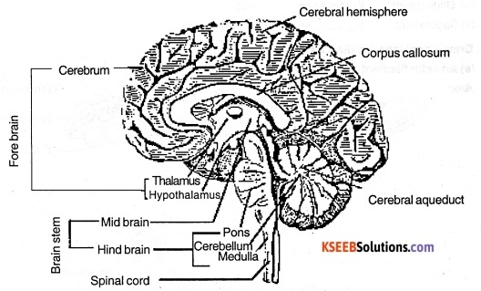
Forebrain:
The forebrain consists of cerebrum, thalamus and hypothalamus. Cerebrum is divided longitudinally into two halves, left and right cerebral hemispheres. The hemispheres are connected by a tract of nerve fibres called corpus callosum. The layer of cells which covers the cerebral hemisphere is called cerebral cortex and is thrown into prominent folds. It is referred to as grey matter due to greyish appearance. The cerebral cortex contains motor areas, sensory areas and association areas which are responsible for complex functions like intersensory associations, memory and communication.
Fibres of the tracts are covered with the myelin sheath, which constitute the inner part of cerebral hemisphere. This layer is white in colour, hence called white matter. The cerebrum wraps around a structure called thalamus, which is a major co-ordinating centre for sensory and motor signalling. Hypothalamus lies at the base of the thalamus and contains several group of neurosecretory cells, which secrete hormones called hypothalamic hormones. The inner parts of cerebral hemispheres and a group of associated deep structures like amygdala, hippocampus, etc. form a complex structure called the limbic lobe or limbic system.
![]()
(ii) Mid brain:
The midbrain is located between the thalamus / hypothalamus of the forebrain and pons of the hind brain. A canal called the cerebral aqueduct passes through the mid brain. The dorsal portion of the midbrain consists of four round swellings (lobes) called Corpora quadrigemina. Midbrain and hindbrain form the brain stem.
(iii) Hindbrain:
Hindbrain comprises pons, cerebellum and medulla. Pons consists of fibre tracts that interconnect different regions of the brain. Cerebellum has very convoluted surface in order to provide the additional space for many more neurons. The medulla of the brain is connected to the spinal cord.
(b) Eye:
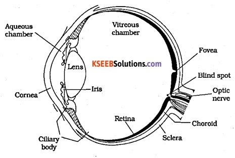
The adult human eye ball is nearly a spherical structure. The wall of the eye ball is composed of three layers. The external layer called Sclera is composed of a dense connective tissue. The middle layer choroid contains many blood vessels and looks bluish in colour. The choroid layer is thin over the posterior two – thirds of the eyeball, but it becomes thick in the anterior part to form the ciliary body. The ciliary body continues to form a pigmented and opaque structure called the iris.
The eyeball contains a transparent crystalline lens which is held in place by ligaments attached to the ciliary body. The aperture surrounded by the iris is called the pupil. The diameter of the pupil is regulated by the muscle fibre of iris. The inner layer is the retina and it contains three layers of cells called ganglion cells, bipolar cells and photoreceptor cells. Rods and cones are two types of photoreceptor cells. The optic nerves leave the eye and the retinal blood vessels enter it at a point medial to and slightly above the posterior pole of the eye ball. Photoreceptor cells are not present in that region and hence it is called the blind spot.
At the posterior pole of the eye lateral to the blind spot, there is a yellowish pigmented spot called macula lutea with a central pit called the fovea. The fovea is a thinned out portion of the retina where only the cones are densely packed. It is the point where the visual activity is the greatest.
The space between the cornea and the lens is called the aqueous chamber and contains a thin watery fluid called aqueous humor. The space between the lens and the retina is called the vitreous chamber and is filled with a transparent gel called vitreous humor.
(c) Ear
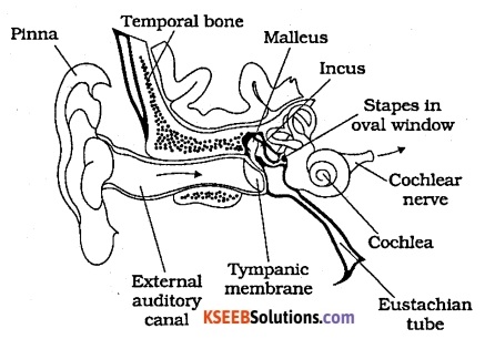
Ear is divided into three major sections called the outer ear, the middle ear and the inner ear. The outer ear consists of the pinna and external auditory meatus. The external auditory meatus leads inwards and extends up to the tympanic membrane (ear drum). There are very fine hairs and wax- secreting sebaceous glands in the skin of the pinna and the meatus. The tympanic membrane is composed of connective tissues.
Covered with skin outside and with mucus membrane inside. The middle ear contains three ossicles called malleus, incus and stapes which are attached to one another in a chain-like fashion. The malleus is attached to the tympanic membrane and the stapes is attached to the oval window of the cochlea. An Eustachian tube connects the middle ear cavity with the pharynx.
The fluid filled inner ear called labyrinth consists of two parts, the bony and the membranous labyrinths. The bony labyrinth is a series of channels. Inside these channels lies the membranous labyrinth, which is surrounded by a fluid called perilymph. The membranous labyrinth is filled with a fluid called endolymph. The coiled portion of the labyrinth is called cochlea.

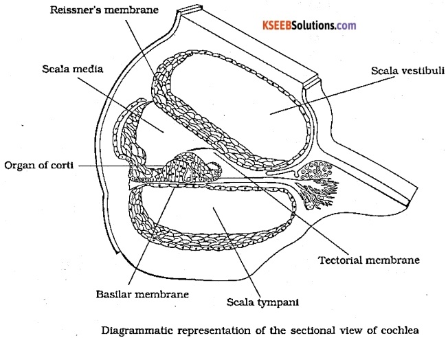
The membranes consisting cochlea, the reissners and basilar, divide the surrounding perilymph filled bony labyrinth into an upper scala vestibuli and a lower scala tympani. The space within cochlea called scala media is filled with endolymph. At the base of the cochlea, the scala vestibuli ends at the oval window, while the scala tympani terminates at the round window which opens to the middle ear.
The organ of Corti is a structure located on the basilar membrane which contains hair cells that act as auditory receptors. The hair cells are present in rows on the internal side of the organ of Corti. The basal end of the hair ceil is in close contact with the afferent nerve fibres. A large number of processes called stereo cilia are projected from the apical part of each hair cell.
Above the rows of the hair cells is a thin elastic membrane called tectorial membrane. The inner ear also contains a complex system called vestibular apparatus, located above the cochlea. The vestibular apparatus is composed of three semicircular canals and the otolith organ consisting of the saccule and utricle. Each semi circular canal lies in a different plane at right angles to each other. The membranous canals are suspended in the perilyrrph of the bony canals. The base of canals is swollen and is called ampulla, which contains a projecting ridge called crista ampullaris which has hair cells. The saccule and utricle contain a projecting ridge called macula.
![]()
Question 2.
Compare the following:
(a) Central neural system (CNS) and Peripheral neural system (PNS)
(b) Resting potential and action potential
(c) Choroid and retina
Answer:
(a) The central neural system (CNS) includes the brain and the spinal cord and is the – site of information processing and control. The PNS comprises of all the nerves of the body associated with the CNS (brain and spinal cord)
(b) Resting potential is the electrical potential difference across the resting plasma membrane. Here the outer surface of the axonal membrane is positively charged and inner surface is negatively charged and therefore is polarised.
The electrical potential difference across the plasma membrane, when the membrane is depolarised is called the action potential. Here the outer membrane is negatively charged and inner membrane is positively charged.
(c) Choroid layer is the middle layer of eye and is brownish black in colour. It is highly vascular and have pigments known as melanocytes which give colour to eye. Retina is the inner most and incomplete layer that extends up to ciliary body. The outer most layer of retina has two types of cells namely rods and cones.
Question 3.
Explain the following processes:
(a) Polarisation of the membrane of a nerve fibre
(b) Depolarisation of the membrane of a nerve fibre
(c) Conduction of a nerve impulse along a nerve fibre
(d) Transmission of a nerve impulse across a chemical synapse
Answer:
(a) When a neuron is not conducting any impulse; i.e resting, the axoplasm inside the axon contains high concentration of K+ and negatively charged proteins and low concentration of Na+. In contrast, the fluid outside the axon contains a low concentration of K+, a high concentration of Na+ and thus form a concentration gradient. These ionic gradients across the resting membrane are maintained by the active transport of ions by the sodium-potassium pump which transports 3 Na+ outwards for 2K+ into the cell. As a result, the outer surface of the axonal membrane possesses a positive charge while its inner surface becomes negatively charged and therefore is polarised.
(b) When a stimulus is applied at a site, (eg: site A) on the polarised membrane, the membrane at the site A becomes freely permeable to Na+. This leads to a rapid influx of Na+ followed by the reversal of the polarity at that site, i.e the outer surface of the membrane becomes negatively charged and the inner side becomes positively charged. The polarity of the membrane at the site A is thus reversed and hence depolarised.
(c)

Consider site A to be the site of excitation and hence depolarised. At sites immediately ahead, the axon (eg: site B) membrane has a .positive charge on the outer surface and a negative charge on its inner surface. As a result, a current flows on the inner surface from site A to site B. On the outer surface current flows from site B to site A to complete the circuit of current flow. Hence, the polarity at the site is reversed, and an action potential is generated at site B. Thus, the impulse generated at site A arrives at site B. The sequence is repeated along the length of the axon and consequently the impulse is conducted.
![]()
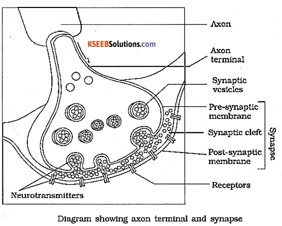
(d) A nerve impulse is transmitted from one neuron to another through junctions called synapses. At a chemical synapse, the membranes of the pre – and post – synaptic neurons are separated by a fluid-filled space called Synaptic cleft. Chemicals called neurotransmitters are involved in the transmission of impulses at these synapses. When an impulse arrives at the axon terminal, it stimulates the movement of the synaptic vesicles towards the membrane where they fuse with the plasma membrane and release their neurotransmitters in the synaptic cleft.
The released neurotransmitters bind to their specific receptors, present on the post-synaptic membrane. This binding opens ion channels allowing the entry of ions which can generate a new potential in the post-synaptic neuron. The new potential developed may be either excitatory or inhibitory.
Question 4.
Draw labelled diagrams of the following:
(a) Neuron
(b) Brain
(c) Eye
(d) Ear
Answer:
(a) Neuron
(b) Brain
The human brain is well protected by the skull. Inside the skull, the brain is covered by cranial meninges consisting of three layers namely outer durometer, middle layer called arachnoid and inner layer pia mater. The brain is divided into three major parts.
- Forebrain
- Mid brain
- Hindbrain.
Forebrain:
The forebrain consists of cerebrum, thalamus and hypothalamus. Cerebrum is divided longitudinally into two halves, left and right cerebral hemispheres. The hemispheres are connected by a tract of nerve fibres called corpus callosum. The layer of cells which covers the cerebral hemisphere is called cerebral cortex and is thrown into prominent folds. It is referred to as grey matter due to greyish appearance. The cerebral cortex contains motor areas, sensory areas and association areas which are responsible for complex functions like intersensory associations, memory and communication.
Fibres of the tracts are covered with the myelin sheath, which constitute the inner part of cerebral hemisphere. This layer is white in colour, hence called white matter. The cerebrum wraps around a structure called thalamus, which is a major co-ordinating centre for sensory and motor signalling. Hypothalamus lies at the base of the thalamus and contains several group of neurosecretory cells, which secrete hormones called hypothalamic hormones. The inner parts of cerebral hemispheres and a group of associated deep structures like amygdala, hippocampus, etc. form a complex structure called the limbic lobe or limbic system.
(ii) Mid brain:
The midbrain is located between the thalamus / hypothalamus of the forebrain and pons of the hind brain. A canal called the cerebral aqueduct passes through the mid brain. The dorsal portion of the midbrain consists of four round swellings (lobes) called Corpora quadrigemina. Midbrain and hindbrain form the brain stem.
(iii) Hindbrain:
Hindbrain comprises pons, cerebellum and medulla. Pons consists of fibre tracts that interconnect different regions of the brain. Cerebellum has very convoluted surface in order to provide the additional space for many more neurons. The medulla of the brain is connected to the spinal cord.
(c) Eye:

The adult human eye ball is nearly a spherical structure. The wall of the eye ball is composed of three layers. The external layer called Sclera is composed of a dense connective tissue. The middle layer choroid contains many blood vessels and looks bluish in colour. The choroid layer is thin over the posterior two – thirds of the eyeball, but it becomes thick in the anterior part to form the ciliary body. The ciliary body continues to form a pigmented and opaque structure called the iris.
The eyeball contains a transparent crystalline lens which is held in place by ligaments attached to the ciliary body. The aperture surrounded by the iris is called the pupil. The diameter of the pupil is regulated by the muscle fibre of iris. The inner layer is the retina and it contains three layers of cells called ganglion cells, bipolar cells and photoreceptor cells. Rods and cones are two types of photoreceptor cells. The optic nerves leave the eye and the retinal blood vessels enter it at a point medial to and slightly above the posterior pole of the eye ball. Photoreceptor cells are not present in that region and hence it is called the blind spot.
At the posterior pole of the eye lateral to the blind spot, there is a yellowish pigmented spot called macula lutea with a central pit called the fovea. The fovea is a thinned out portion of the retina where only the cones are densely packed. It is the point where the visual activity is the greatest.
The space between the cornea and the lens is called the aqueous chamber and contains a thin watery fluid called aqueous humor. The space between the lens and the retina is called the vitreous chamber and is filled with a transparent gel called vitreous humor.
![]()
(d) Ear
Ear is divided into three major sections called the outer ear, the middle ear and the inner ear. The outer ear consists of the pinna and external auditory meatus. The external auditory meatus leads inwards and extends up to the tympanic membrane (ear drum). There are very fine hairs and wax- secreting sebaceous glands in the skin of the pinna and the meatus. The tympanic membrane is composed of connective tissues.
Covered with skin outside and with mucus membrane inside. The middle ear contains three ossicles called malleus, incus and stapes which are attached to one another in a chain-like fashion. The malleus is attached to the tympanic membrane and the stapes is attached to the oval window of the cochlea. An Eustachian tube connects the middle ear cavity with the pharynx.
The fluid filled inner ear called labyrinth consists of two parts, the bony and the membranous labyrinths. The bony labyrinth is a series of channels. Inside these channels lies the membranous labyrinth, which is surrounded by a fluid called perilymph. The membranous labyrinth is filled with a fluid called endolymph. The coiled portion of the labyrinth is called cochlea.
The membranes consisting cochlea, the reissners and basilar, divide the surrounding perilymph filled bony labyrinth into an upper scala vestibuli and a lower scala tympani. The space within cochlea called scala media is filled with endolymph. At the base of the cochlea, the scala vestibuli ends at the oval window, while the scala tympani terminates at the round window which opens to the middle ear.
The organ of Corti is a structure located on the basilar membrane which contains hair cells that act as auditory receptors. The hair cells are present in rows on the internal side of the organ of Corti. The basal end of the hair ceil is in close contact with the afferent nerve fibres. A large number of processes called stereo cilia are projected from the apical part of each hair cell.
Above the rows of the hair cells is a thin elastic membrane called tectorial membrane. The inner ear also contains a complex system called vestibular apparatus, located above the cochlea. The vestibular apparatus is composed of three semicircular canals and the otolith organ consisting of the saccule and utricle. Each semi circular canal lies in a different plane at right angles to each other. The membranous canals are suspended in the perilyrrph of the bony canals. The base of canals is swollen and is called ampulla, which contains a projecting ridge called crista ampullaris which has hair cells. The saccule and utricle contain a projecting ridge called macula.
Question 5.
Write short notes on the following:
(a) Neural coordination
(b) Forebrain
(c) Midbrain
(d) Hindbrain
(d) Retina
(f) Ear ossicles
(g) Cochlea
(h) Organ of Cortl
(I) Synapse
Answer:
(a) Neural co-ordination: Co-ordination is the process through which two or more organs interact and complement the functions of one another. In our body the neural system and the endocrine system jointly co-ordinate and integrate all the activities of the organs so that they function in a synchronised fashion. The neural system provides an organised network of point to point connections for a quick co-ordinations. Neural – co-ordination includes mechanisms like transmission of nerve impulse, impulse conduction across a synapse and reflex action.
(b) Forebrain:
The forebrain consists of cerebrum, thalamus and hypothalamus. Cerebrum is divided longitudinally into two halves, left and right cerebral hemispheres. The hemispheres are connected by a tract of nerve fibres called corpus callosum. The layer of cells which covers the cerebral hemisphere is called cerebral cortex and is thrown into prominent folds. It is referred to as grey matter due to greyish appearance. The cerebral cortex contains motor areas, sensory areas and association areas which are responsible for complex functions like intersensory associations, memory and communication.
Fibres of the tracts are covered with the myelin sheath, which constitute the inner part of cerebral hemisphere. This layer is white in colour, hence called white matter. The cerebrum wraps around a structure called thalamus, which is a major co-ordinating centre for sensory and motor signalling. Hypothalamus lies at the base of the thalamus and contains several group of neurosecretory cells, which secrete hormones called hypothalamic hormones. The inner parts of cerebral hemispheres and a group of associated deep structures like amygdala, hippocampus, etc. form a complex structure called the limbic lobe or limbic system.
(c) Midbrain:
The midbrain is located between the thalamus / hypothalamus of the forebrain and pons of the hind brain. A canal called the cerebral aqueduct passes through the mid brain. The dorsal portion of the midbrain consists of four round swellings (lobes) called Corpora quadrigemina. Midbrain and hindbrain form the brain stem
(d) Hindbrain:
Hindbrain comprises pons, cerebellum and medulla. Pons consists of fibre tracts that interconnect different regions of the brain. Cerebellum has very convoluted surface in order to provide the additional space for many more neurons. The medulla of the brain is connected to the spinal cord.
(e) Retina: Retina is the inner layer of the eye and contains three layers of cells; ganglion cells, bipolar cells and photoreceptor cells, (from inside to outside respectively). There are two types of photoreceptor cells, namely rods and cones. These cells contain the light sensitive proteins called the photo pigments. The daylight vision and colour vision are functions of cones and the twilight vision is the function of the rods. The rods contain a purplish – red protein called the rhodopsin or visual purple, which contains a derivative of Vitamin A. There are three types of cones that respond to red, green and blue lights.
(f) Ear ossicles: The ear ossicles seen in the middle ear are malleus, incus and stapes. They are attached to one another in a chain like fashion. Malleus is attached to the tympanic membrane and the stapes is attached to the oval window of the cochlea. The ear ossicles increase the efficiency of transmission of sound waves to the inner ear.
(g) Cochlea: The coiled portion of the labyrinth is called cochlea. The membranes constituting cochlea, the reissner’s and basilar, divide the surrounding perilymph filled bony labyrinth into an upper scala vestibuli and a lower scala tympani. The space within cochlea called scala media is filled with endolymph. At the base of the cochlea, the scala vestibuli ends at the oval window, while the scala tympani terminates at the round window which opens to the middle ear.
(h) Organ of Corti: The organ of Corti is a structure located on the basilar membrane which contains hair cells that act as auditory receptors. The hair cells are present in rows on the internal side of the organ of Corti. The basal end of the hair cell is in close contact with the afferent nerve fibres. A large number of processes called stereo cilia are projected from the apical part of each hair cell. Above the rows of the hair cells is a thin elastic membrane called tectorial membrane.
![]()
(i) Synapse: A nerve impulse is transmitted from one neuron to another through junctions called synapses. A synapse is formed by the membranes of a pre- synaptic neuron and a post – synaptic neuron, which may or may not be separated by a gap called synaptic cleft. There are two types of synapses, namely, electrical synapses, the membranes of pre and post synaptic neurons are in very close proximity. Electrical current can flow directly from one neuron into the other across these synapses.
Transmission of an impulse across electrical synapses is very similar to impulse conduction along a single axon. Impulse transmission across an electrical synapse is always faster than that across a chemical synapse. At a chemical synapse, the membranes of the pre and post synaptic neurons are separated by a fluid – filled space called synaptic cleft
Question 6.
Give a brief account of:
(a) Mechanism of synaptic transmission
(b) Mechanism of vision
(c) Mechanism of hearing
Answer:
(a) Mechanism of synaptic transmission: When an impulse reaches at the axon terminal, it stimulates the movement of the synaptic vesicles towards the synaptic cleft and releases their neurotransmitters. The neurotransmitter binds with the receptors found on the post synaptic membrane. This binding opens ion channels allowing the entry of ions which can generate an action potential on the post synaptic neuron.
(b) Mechanism of vision: The rays in visible wavelength focussed on the retina through the cornea and lens generate potentials (impulse) in rods and cones. The photo- sensitive compounds (photo pigments) in the human eyes is composed of opsin and retinal. Light induces dissociation of the retinal from opsin resulting in changes in the structure of the opsin. This causes membrane permeability to change.
As a result, potential differences are generated in the photoreceptor cells. This produces a signal that generates action potentials in the ganglion cells through the bipolar cells. These action potentials are transmitted by the optic nerves to the visual cortex area of the brain, where the neural impulses are analysed and the image formed on the retina is recognised based on earlier memory and experience.
![]()
(c) Mechanism of hearing: The external ear receives sound waves and directs them to the ear drum. The ear drum vibrates in response to the sound waves and these vibrations are transmitted through the ear ossicles to the oval window. The vibrations are passed through the oval window on to the fluid of the cochlea, where they generate waves in the lymphs.
The waves in the lymphs induce a ripple in the basilar membrane. These movements of the basilar membrane bend the hair cells, pressing them against the tectorial membrane. As a result, nerve impulses are generated in the associated afferent neurons. These impulses are transmitted by the afferent fibres via auditory nerves to the auditory cortex of the brain, where the impulses are analysed and the sound is recognised.
Question 7.
Answer briefly:
(a) How do you perceive the colour of an object?
(b) Which part of our body helps us in maintaining the body balance?
(c) How does the eye regulate the amount of light that falls on the retina.
Answer:
(a) Rods and cones are photoreceptor cells that contain light sensitive proteins called photo pigments. The daylight vision and colour vision are functions of cones. There are three types of cones which respond to red, green and blue lights. The sensations of different colours are produced by various combinations of these cones and their photo pigments. When these cones are stimulated equally, a sensation of white light is produced.
(b) The crista and macula are the specific receptors of the vestibular apparatus responsible for maintenance of balance of the body.
(c) The pupil in the eye functions like an aperture. This dilates in case of low light and constricts in case of intense light thereby regulating the amount of light falling on the retina.
Question 8.
Explain the following:
(a) Role of Na+ in the generation of action potential.
(b) Mechanism of generation of light-induced impulse in the retina.
(c) Mechanism through which a sound produces a nerve impulse in the inner ear.
Answer:
(a) When a stimulus is applied at a site, (eg: site A) on the polarised membrane, the membrane at the site A becomes freely permeable to Na+ . This leads to a rapid influx of Na+ followed by the reversal of the polarity at that site, i.e., the outer surface of the membrane becomes negatively charged and the inner side becomes positively charged. The polarity of the membrane at the site A is thus reversed and hence depolarised. Thus action potential is generated across the plasma membrane.
(b) Mechanism of vision: The rays in visible wavelength focussed on the retina through the cornea and lens generate potentials (impulse) in rods and cones. The photo- sensitive compounds (photo pigments) in the human eyes is composed of opsin and retinal. Light induces dissociation of the retinal from opsin resulting in changes in the structure of the opsin. This causes membrane permeability to change.
As a result, potential differences are generated in the photoreceptor cells. This produces a signal that generates action potentials in the ganglion cells through the bipolar cells. These action potentials are transmitted by the optic nerves to the visual cortex area of the brain, where the neural impulses are analysed and the image formed on the retina is recognised based on earlier memory and experience.
(c) Mechanism of hearing: The external ear receives sound waves and directs them to the eardrum. The eardrum vibrates in response to the sound waves and these vibrations are transmitted through the ear ossicles to the oval window. The vibrations are passed through the oval window on to the fluid of the cochlea, where they generate waves in the lymphs.
The waves in the lymphs induce a ripple in the basilar membrane. These movements of the basilar membrane bend the hair cells, pressing them against the tectorial membrane. As a result, nerve impulses are generated in the associated afferent neurons. These impulses are transmitted by the afferent fibres via auditory nerves to the auditory cortex of the brain, where the impulses are analysed and the sound is recognised.
Question 9.
Differentiate between:
(a) Myelinated and non-myelinated axons
(b) Dendrites and axons
(c) Rods and cones
(d) Thalamus and Hypothalamus
(e) Cerebrum and Cerebellum
Answer:
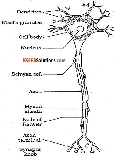
(a) The myelinated nerve fibres are enveloped with Schwann cells, which form a myelin sheath around the axon. The gaps between two adjacent myelin sheaths are called nodes of Ranvier. Myelinated nerve fibres are found in spinal and cranial nerves.
Unmyelinated nerve fibre is enclosed by a Schwann cell that does not form a myelin sheath around the axon, and is commonly found in autonomous and the somatic nervous systems.
![]()
(b) Dendrites are short fibres which branch repeatedly and project out of the cell body. It also contains Nissl’s granules. These fibres transmit impulses towards the cell body. The axon is a long fibre, the distal and which is branched. Each branch terminates as a bulb like structure called synaptic knob which possess synaptic vesicles containing chemicals called neurotransmitters. The axon transmit nerve impulses away from the cell body to a synapse orto a neuro-muscular junction.
(c)
| Rods | Cones |
| (i) Responsible for twilight vision. | (i) Responsible for daylight and colour vision. |
| (ii) More in number | (ii) Less in number |
| (iii) Respond to lower light intensity. | (iii) Sensitive to bright light |
| (iv) Absent in fovea centralis | (iv) Present in fovea |
| (v) Contain pigment Rhodopsin | (v) Contain pigment iodopsin |
| (v) Contain pigment | (v) Contain pigment |
(d) The cerebrum wraps around a structure called thalamus, which is major coordinating centre for sensory and motor signaling. It functions as a relay station. Hypothalamus lies at the base of thalamus. This portion contains a number of centres which control body temperature, urge for eating and drinking. It also contains several groups of neuro secretory cells, which secrete hormones called hypothalamic hormones.
(e)
| Cerebrum | Cerebellum |
| (i) Portion of fore brain. (ii) Seat of highest mental faculties, governs reasoning, learning, memory, intelligence (iii) Responds to heat, cold, pain, touch, light and pressure. | (i) Portion of hind brain (ii) Regulates and coordinates the contraction of skeletal muscles. (iii) Maintains equilibrium and controls posture. |
Question 10.
Answer the following:
(a) Which part of the ear determines the pitch of a sound?
(b) Which part of the human brain is the most developed?
(c) Which part of our central neural system acts as a master clock?
Answer:
(a) Organ of Corti
(b) Cerebrum
(c) Pineal gland
Question 11.
The region of the vertebrate eye, where the optic nerve passes out of the retina, is called the ………………….
(a) fovea
(b) Iris
(c) blind spot
(d) optic chiasma
Answer:
(c) blind spot
Question 12.
Distinguish between:
(a) afferent neurons and efferent neurons
(b) impulse conduction in a myelinated nerve fibre and unmyelinated nerve fibre
(c) aqueous humor and vitreous humor
(d) blind spot and yellow spot
(f) cranial nerves and spinal nerves.
Answer:
(a) The neuron which connects sense organs aw’ brain is called afferent neuron. The neuron which connects brain and effector organs or the concerned peripheral tissues organs is called efferent neuron.
(b) In a myelinated neuron, impulse is transmitted by chemical method. It allows fast, saltatory movement of action potentials from node to node. In an unmyelinated neuron, impulse is transmitted by clerical mechanism. The conduction velocity of myelinated neurons vary linearly with axon diameter, whereas the speed of unmyelinated neurons vary roughly as the square root of the diameter of axons. Myelinated axon conduction is fast and energy efficient.
(c) The space between the cornea and the lens is called the aqueous chamber and contains a thin watery fluid called aqueous humor. The space between the lens and the retina is called the vitreous chamber and is filled with a transparent gel called vitreous humor.
(d) The optic nerves leave the eye and the retinal blood vessels enter it at a point medial to and slightly above the posterior pole of the eyeball called the blind spot. It doesn’t contain any photoreceptor cells (rods and cones). At the posterior pole of the eye lateral to the blind spot, is a yellowish pigmented spot called macula lutea (yellow spot). It contains cones only and the point where the visual acuity is the greatest
(e) Cranial nerves are the nerves that emerge directly from the brain stem in contrast to spinal nerves which emerge from segments of the spinal cord.
1st PUC Biology Neural Control and Coordination Additional Questions and Answers
1st PUC Biology Neural Control and Coordination One Mark Questions
Question 1.
What are meninges? (July 2011)
Answer:
The connective tissue membranes around brain are called meninges.
Question 2.
What is CNS?
Answer:
CNS or central nervous system is that includes brain and spinal cord.
Question 3.
What is Durometer?
Answer:
The outermost layer of meninges of CNS is Durometer.
Question 4.
What is corpus callosum?
Answer:
A transverse band of myelinated nerve fibers that connects cerebral hemispheres is called corpus callosum.
Question 5.
What is reflex arc? (June 2009)
Answer:
The path through which the electrochemical impulses are generated in response to a stimulus is called reflex arc.
Question 6.
What is reflex action?
Answer:
The quick, spontaneous and involuntary action induced by the nervous system in
response to a stimulus is called reflex action.
Question 7.
Which part of brain maintains body equilibrium? (Oct. 2000)
Answer:
Cerebellum is the centre for muscular coordination and equilibrium.
Question 8.
Name the protective covering of the brain. (April 2001)
Answer:
Meninges
Question 9.
Name the deep bridge of rieVve fibres which Joins cerebral hemispheres*. (Oct. 2003)
Answer:
Corpus callosum.
Question 10.
Name the structural and functional unit of nervous system. (July 2006)
Answer:
neuron
Question 11.
What is meant by co-ordination in our body?
Answer:
Co-ordination is the process through which two or more organs interact and complement the functions of one another.
Question 12.
What are afferent nerve fibers?
Answer:
The nerve fibres that transmit impulses from tissues / organs to the CNS are called afferent nerve fibres.
Question 13.
What are efferent fibres ?
Answer:
The nerve fibres that transmit regulatory impulses from the CNS to the concerned peripheral tissues / organs are called efferent nerve fibres.
Question 14.
Name the classification of autonomic neural system.
Answer:
- Sympathetic neural system
- Parasympathetic neural system
Question 15.
What are the major parts of a neuron ?
Answer:
Neuron is composed of cell body, dendrites and axon.
Question 16.
What are Nisei’s granules?
Answer:
Mr.’Nissl’s granules are certain granular bod¬ies present in the cytoplasm of the cell body of a neuron.
Question 17.
What are dendrites?
Answer:
Short fibres that branch repeatedly and project out of the cell body and also contain Nissl’s granules are called dendrites.
Question 18.
Where are neuro transmitters found ?
Answer:
Neurotransmitters are chemicals found in synaptic vesicles present in synaptic knob.
Question 19.
Name the three types of neurons.
Answer:
Multipolar, bipolar and unipolar neurons.
Question 20.
Where are multipolar neurons found in human body?
Answer:
Cerebral cortex …
Question 21.
What are nodes of Ranvier ?
Answer:
The gaps between two adjacent myelin sheaths of axon are called nodes of Ranvier.
Question 22.
Where exactly are synaptic vesicles located? What is their role ? (Foreign 2006)
Answer:
Synaptic vesicles are found in synaptic knob. They contain chemicals called neurotransmitters which are involved in transmission of impulses.
Question 23.
What are the charges on outer surface and Inner surface of axonal membrane during rest?
Answer:
Outer surface is positively charged and inner surface is negatively charged.
Question 24.
What are the functions of association areas?
Answer:
Association areas are responsible for complex functions like inter sensory associations memory and communication.
![]()
Question 25.
Write one function of limbic system in human brain. (Foreign 2006)
Answer:
Sexual behaviour, excitement, pleasure, rage, fear and motivation.
Question 26.
What is the canal passing through mid brain called?
Answer:
Cerebral aqueduct.
Question 27.
What is corpora quadrlgemina ?
Answer:
Corpora quadrigemina are four round swellings (lobes) found in the dorsal portion of midbrain.
Question 28.
Name the three regions of hindbrain.
Answer:
Pons, cerebellum and medulla.
Question 29.
What is the function of medulla oblongata.
Answer:
It contains centres which control respiration, cardiovascular reflexes and gastric secretions.
Question 30.
Which is the visible coloured portion of the eye ?
Answer:
Iris.
Question 31.
Name the external layer of the eye.
Answer:
Sclera
Question 32.
Name the area of retina which contains only cones and no rods. (Delhi 1997)
Answer:
Fovea
Question 33.
Why is blind spot devoid of the ability for vision?
Answer:
Blind spot does not contain any photoreceptor cells (rods and cones), hence devoid of vision.
Question 34.
Name the photosensitive compounds in human eye.
Answer:
Mropsin and retinal.
Question 35.
Name the fluid in which the membranous labyrinth of the inner ear floats. (Delhi 1997)
Answer:
Perilymph
Question 36.
Give the technical names of the auditory ossicles in their natural sequence. (All India 1997)
Answer:
Malleus, incus and stapes.
Question 37.
What connects the middle ear cavity with the pharynx?
Answer:
Eustachian tube.
Question 38.
Name the space within cochlea filled with endolymph.
Answer:
Scala media.
![]()
Question 39.
What does vestibular apparatus composed of?
Answer:
Three semicircular canals and otolith organ.
Question 40.
What is macula?
Answer:
The saccule and utricle contain a projecting ridge called macula.
Question 41.
Name the receptors responsible for body balance and posture.
Answer:
Crista and macula.
Question 42.
What is the function of Eustachian tube?
Answer:
It helps in equalising the pressures on either sides of the ear drum.
1st PUC Biology Neural Control and Coordination Two Marks Questions
Question 1.
What is reflex arc and reflex action.
Answer:
The path through which the electro-chemical impulses are generated in response to a stimulus is called reflex arc. It is the simplest functional unit of the nervous system by which an impulse produces a reflex action.The quick, spontaneous and involuntary action induced by the nervous system in response to a stimulus is called reflex action.
Question 2.
What are the functions of CSF.
Answer:
- It acts as a buffer, protecting the brain and spinal cord.
- It conveys nourishment to the tissues of the brain and spinal cord.
- It protects both brain and spinal cord from shocks and maintains constant pressure with in cranium.
Question 3.
Write the functions of hypothalamus.
Answer:
- Thermo regulation
- Biological clock system
- Autonomic nervous system control
- Sleep
- Controlling body temperature, appetite, secretion of pituitary.
Question 4.
What are Meninges? Mention different layers of Meninges.
Answer:
The connective tissue membranes around brain are called meninges. They protect the delicate nerve structure, carry the blood vessels to it and by the secretion of a fluid
(C S F) minimize any blow or concussion. There are three layers of Meninges;
- Durometer
- The arachnoid
- Piamater
Question 5.
What happens if the cerebellum is damaged. (Oct. 97)
Answer:
As the cerebellum is concerned with the maintenance of equilibrium of the body and coordination of contractions and relaxations of muscles, a damage to the, cerebellum leads to loss of the ability in muscular co-ordination and equilibrium in a person.
![]()
Question 6.
List any four functions of hind brain. (July 06)
Answer:
The functions of the hind brain are
- It regulates posture and postural activities.
- It plays an important part in muscular co ordination and maintenance of balance
- Cerebellar hemisphere controls muscle tone and posture on its side
- It regulates smooth and precise goal oriented movements.
Question 7.
Briefly give an account of somatic and autonomic nervous system.
Answer:
The PNS is divided into two divisions:
- Somatic neural system
- Autonomic neural system (ANS)
Somatic neural system relays impulses from the CNS to skeletal muscles while the autonomic neural system transmits impulses from the CNS to the involuntary organs and smooth muscles of the body. The ANS is further classified into sympathetic neural system and parasympathetic neural system.
Question 8.
What are the functions of hypothalamus?
Answer:
- It controls body temperature, urge for eating and drinking.
- It contains cells that secrete hormones called hypothalamic hormones.
- It is involved in the regulation of sexual behaviour, expression, emotional reactions and motivation.
Question 9.
Write the difference between cerebrum and cerebellum
Answer:
| Cerebrum | Cerebellum |
| (a) It is a part of forebrain (b) It is meant for memory, intelligence and control of voluntary movements | (a) It is a part of hindbrain (b) It controls the movements and help in maintaining body posture. |
Question 10.
Why are gray matter and white matter contained In human nervous system name so? (Delhi 2006)
Answer:
Grey matter:
It contains neural cell bodies (spindle, pyramidal and stellate neurons) which give them greyish appearance.
White matter:
This region contains millions of axons with myelin sheath which gives them an opaque white appearance.
Question 11.
Name and differentiate between two types of synapses. (Foreign 2004)
Answer:
The two types of synapses present are:
- Electrical synapse
- Chemical synapse
| Electical synapse | Chemical synapse |
| (a) Electrical current flows directly from one neuron to other. (b) Synaptic cleft is very narrow (c) Impulse transmission is faster (d) These are less common | (a) Signal transmissions involve chemical called neurotransmitters. (b) Synaptic cleft is wider. (c) Impulse transmission is slower. (d) These are more common. |
Question 12.
Name the ear ossicles in the order of arrangement in humans. What role do they play in hearing?
Answer:
Malleus, Incus, and Stapes are the ear ossicles. The vibrations due to sound waves are transmitted through the ear ossicles to the oval window which are further transmitted to endolymph.
Question 13.
What is blind spot? Why is it so named? (Foreign 2003)
Answer:
The point where optic nerves leave the eye and the retinal blood vessels enter which is slightly above posterior pole of the eye ball is called blind spot. It lacks photoreceptor cells (rods and cones) and is devoid of vision. Hence called blind spot.
Question 14.
What happens when the membrane of a nerve cell carries out a sodium pump? (Delhi 2001)
Answer:
When the membrane of a nerve cell carries out a sodium pump, it transports 3 Na+ ions outwards for 2 K+ ions into the cell. As a result, the outer surface of the axonal membrane posses a positive charge while its inner surface is negatively charged and therefore polarised.
Question 15.
What are the events that take place at the point of simulation of an axon? (Delhi 1997)
Answer:
When a stimulus is applied at a site on the polarised membrane, it becomes freely permeable to Na+. This leads to a rapid influx of Na+ which causes the outer surface of the membrane to be negatively charged and the inner side becomes positively charged i.e., depolarisation takes place.
Question 16.
What constitutes the outer ear. Mention one function of each.
Answer:
The outer ear consists of the pinna and external auditory meatus.
- Pinna collects the vibrations in the air which produce sound.
- There are very fine hairs and wax secreting sebaceous glands in the skin of pinna and the meatus.
![]()
1st PUC Biology Neural Control and Coordination Three Marks Questions
Question 1.
Draw a diagram to show the path followed by the nerve impulse from the receptor to the effector in a spinal reflex arc. Label any six parts. (All India 2004 )
Answer:

Question 2.
Where are synaptic vesicles found? Name their chemical contents. What Is the function of these contents?
Answer:
Synaptic vesicles are found in a bulb like structure called synaptic knob. Synaptic vesicles contain chemicals called neurotransmitters. Neurotransmitters are involved in the transmission of impulses at the synapses. They bind to the receptors present on the post – synaptic membrane and generate new potential.
Question 3.
Explain briefly the structure and function of human middle ear. (All India 1997 C)
Answer:
The middle ear contains three ossicles called malleus, incus and stapes which are attached to one another in a chain like fashion. The malleus is attached to the tympanic membrane and the stapes is attached to the oval window of the cochlea. An Eustachian tube connects the middle ear cavity with the pharynx.
Functions:
- The ear ossicles increase the efficiency of transmission of sound waves to the inner ear.
- Eustrachian tube helps in equalising the pressures on either sides of the ear drum.
Question 4.
Name the three types of neurons based on the number of axon and dendrites and give the location of their presence.
Answer:
(i) Unipolar neurons:
- Cell body with one axon.
- found in embryonic stage
(ii) Bipolar neurons:
- Cell body with one axon and one dendrite
- found in retina of the eye
(iii) Multipolar neurons:
- Cell body with one axon and two or more dendrites
- Found in cerebral cortex
Question 5.
Draw a diagram showing axon terminal and synapse and label any six parts
Answer:
Question 6.
Draw a neat labelled diagram of sectional view of cochlea.
Answer:
1st PUC Biology Neural Control and Coordination Five Marks Questions
Question 1.
List out the functions of different parts of the human brain. (April 97)
Answer:
The human brain has 3 main divisions namely Forebrain, Midbrain and Hindbrain. The Forebrain is composed (or subdivided into) of the cerebrum and diencephalon, the Midbrain is also called the mesencephalon with only one subdivision and the hind brain is composed of two subdivisions namely the cerebellem and medulla oblongata.
The functions of all these parts forming the brain are listed below:
(1) Forebrain (prosencephalon)
- Cerebrum – Is a functional center for intelligence, consciousness, voluntary control, memory.
- Diencephalon- (Thalamus & hypothalamus) – While the thalamus acts as a main relay centre for conducting information, the hypothalamus controls the temperature of the body, appetite, fat metabolism, secretions of pituitary, sleep and the emotional states namely fear and anger.
(2) Midbrain (Mesencephalon) is the control centre for visual and auditory reflexes like adjustment of the ear to sound, pupil reflex, blinking etc.
(3) Hindbrain (Rhombencephalon)
- Cerebellum is the control centre for muscular coordination and equilibrium.
- Pons connects various parts of the brain with one another and contains the respiratory centres
- Medulla oblongata: contains the centres that control the heart beat, blood pressure, respiration and also centres that control swallowing, coughing and vomiting.
Question 2.
(i) A person unconsciously withdraws his hand suddenly with a jerk after touching hot plata- Draw a schematic diagram of the nervous pathway involved in the response.
(ii) What is such a response called?
Answer:
(i)
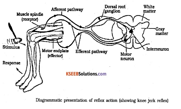
(ii) Such a response is called ‘reflex action’
![]()
Question 3.
Match the following:
I – II
(a) Depolarisation (i) Mid brain
(b) Polarisation (ii) Hind brain
(c) Corpus callosum (iii) Fore brain
(d) Corpora quadrigemina (iv) Action potential
(e) Pons (v) Resting potential
Answer:
(a) → (iv)
(b) → (v)
(c) → (iii)
(d) → (i)
(e) → (ii)
Question 4.
Fill In the blanks.
(a) The………….. fibres transmit impulses from tissues to the CNS and …………..fibres transmit Impulses from CNS to organs.
(b) The gaps between two adjacent myelin sheaths is called …………….
(c) …………….. are responsible for functions like memory and communication.
(d) The coiled portion of the labyrinth is called …………..
(e) ………….. contains hair cells that act as auditory receptors.
Answer:
(a) Afferent, Efferent
(b) Nodes of Ranvier
(c) Association areas
(d) Cochlea
(e) Organ of Corti
Question 5.
State whether the following statements are true / false. Correct the statements If false.
(a) Impulse transmission across electrical synapse is faster than that of chemical
synapse.
(b) During polarisation of axonal membrane, outer surface is negatively charged and inner surface is positively charged.
(c) Middle layer of cranial meninges is called pla mater.
(d) Cerebellum is responsible for controlling respiration and gastric secretions.
(e) Visible coloured portion of the eye Is Iris.
Answer:
(a) True.
(b) False; outer surface is positively charged and inner surface is negatively charged.
(c) False; middle layer is called arachnoid
(d) False; Medulla is responsible for controlling respiration and gastric secretions.
(e) True.