You can Download Chapter 6 Anatomy of Flowering Plants Questions and Answers, 1st PUC Biology Question Bank with Answers, Karnataka State Board Solutions help you to revise complete Syllabus and score more marks in your examinations.
Karnataka 1st PUC Biology Question Bank Chapter 6 Anatomy of Flowering Plants
1st PUC Biology Anatomy of Flowering Plants NCERT Text Book Questions and Answers
Question 1.
State the location and function of different types of meristems.
Answer:
| Types of meristems | Location | Function |
| Apical meristem | Root tips & shoot tips | Growth in length |
| Intercalary meristem | Between mature tissues | repair |
| Secondary meristem | on the periphery of stems & roots | Growth in thickness |
Question 2.
Cork comblum forms tissues that form the cork. Do you agree with this statement? Explain.
Answer:
As the stem continues to increase in girth due to the activity of vascular cambium, the outer cortical and epidermis layers get broken and need to be replaced to provide new protective cell layers. Hence, another meristematic tissue called cork cambium or phellogen develops, usually in the cortex region. Phellogen is a couple of layers thick. It is made of narrow, thin-walled and nearly rectangular cells. The outer cell differentiate into cork while inner cells differentiate into cortex. Thus it is clear how cork cambium forms tissues that form the cork.
Question 3.
Explain the process of secondary growth in the stems of woody angiosperms with the help of schematic diagrams. What is Its significance?
Answer:
The growth of roofs and stems in length with the help of apical meristem is called the primary growth. Most dicotyledonous plants exhibit an increase in girth. This increase is called secondary growth. The tissues involved in secondary growth are the two lateral meristems.
(a) Vascular cambium:
The meristematic layer responsible for cutting off vascular tissues- xylem and phloem is called vascular cambium. In dicot stems, the cells of cambium present between primary xylem and primary phloem is the intrafascicular cambium. The medullary cells adjoining these form intrafascicular cambium. Thus a continuous ring of cambium is formed. Activity of the cambial ring. The cambial ring becomes active and begins to cut off new cells, both towards the inner and the outer sides.
![]()
The cells cut off towards pith, mature into secondary xylem and the cells cut off towards periphery, mature in secondary phloem. The cambium is generally more active on the inner side than on the outer. As a result, the amount of secondary xylem produced is more than secondary phloem and soon forms a compact mass.
The primary and secondary phloems get gradually crushed due to the continued formation and accumulation of secondary xylem. At some places, the cambium forms a narrow band of parenchyma, which passes through the secondary xylem and the secondary phloem in radial directions. These are called medullary rays.
(b) Cork cambium:
As the stem continues to increase in girth due to the activity of vascular cambium, the outer cortical arid epidermis layers get broken and need to be replaced to provide new protective cell layers. Hence, another meristematic tissue called cork cambium or phellogen develops, usually in the cortex region. Phellogen is a couple of layers thick. It is made of narrow, thin-walled and nearly rectangular cells. Phellogen cuts off cells on both sides. The outer cells differentiate into cork while the inner cell differentiates into the secondary cortex. The cork is impervious to water due to suberin deposition in the cell wall.
The cells of the secondary cortex are parenchymatous. Phellogen, phellem and phelloderm are collectively known as periderm. Due to the activity of cork cambium, pressure builds up on the remaining layers peripheral to phellogen and ultimately these layers die and slough off.
At certain regions, the phellogen cuts off closely arranged parenchymatous cells on the outer side instead of cork cell. These parenchymous cells soon rupture the epidermis, forming a lens-shaped openings called lenticels. Lenticels permit the exchange of gases between the outer atmosphere and the internal tissue of the stem.
Question 4.
Draw illustrations to bring out the anatomical difference between
(a) Monocot root and Dicot root
Answer:
(b) Monocot stem and Dicot stem
Answer:
Question 5.
Cut a transverse section of young stem of a plant from your school garden and observe it
Answer:
As shown in the above diagram the vascular bundles in dicot stems are arranged in a ring, while freely are in scattered arrangement in monocot sterns.
Question 6.
The transverse section of a plant material shows the following anatomical features
(a) The vascular bundles are conjoint, scattered and surrounded by a sclerenchymatous bundle sheaths.
(b) Phloem parenchyma is absent. What will you identify It as?
Answer:
It is clear from the above transverse section of the plant that the plant has a monocot stem because vascular bundles are scattered In monocot stems and phloem parenchyma is absent.
![]()
Question 7.
Why are xylem and phloem called complex tissues?
Answer:
Xylem and phloem a composed of several types of cells and they work as a unit. Hence they are called complex tissues.
Question 8.
What is stomatal apparatus? Explain the structure of stomata with a labelled diagram.
Answer:
Stomata are structures present in the epidermis of leaves. Stomata regulate the process transpiration and gaseous exchange. Each stoma is composed of two bean-shaped cells known as guard cells. In grasses, the guard cells are dumbbell-shaped. The outer walls of guard cells are thin and the inner walls are highly thickened.
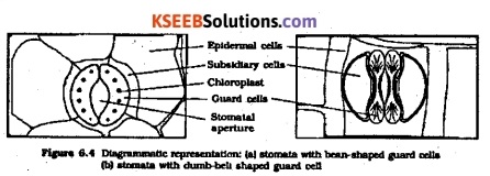
The guard ceils possess chloroplasts and regulate the opening and closing of stomata. Sometimes, a few epidermal cells, in the vicinity of the guard cells become specialised in their shape and size and are known as subsidiary cells. The stomatal aperture, guard ceils and the surrounding subsidiary cells are together called stomatal apparatus
Question 9.
Name the three basic tissue systems in the flowering plants. Give the tissue names under each system.
Answer:
On the basis of their structure and location, there are three types of tissue systems
- Epidermal Tissue system: Epidermis stomata
- Ground or Fundamental Tissue system: Parenchyma, sclerenchyma and collenchyma.
- Vascular or conducting Tissue system: Phloem and Xylem.
Question 10.
How is the study of plant anatomy useful to us?
Answer:
‘The study of plant anatomy is useful in many ways. First of all the study helps us understand the way a plant functions carrying out its routine activities like transpiration, photosynthesis and growth and repair. Second, it helps botanists and agriculture scientists to understand the disease and cure for plants. Plants are important to maintain the ecological balance of the earth, so understanding plant anatomy is a way to understand the large system of the ecology on this planet.
Question 11.
What is periderm? How does periderm formation take place In the dicot stems?
Answer:
As the stem continues to increase in girth due to the activity of vascular cambium, the outer cortical and epidermis layers get broken and need to be replaced to provide new protective cell layers. Hence, sooner or later, another meristematic tissue called cork cambium or phellogen develops, usually in the cortex region. Phellogen is a couple of layers thick. It is made of narrow, thin-walled and nearly rectangular cells.
Phellogen cuts off cells on both sides. The outer cells differentiate into cork or phellem while the inner cells differentiate into secondary cortex or phelloderm. The cork is impervious to water due to suberin deposition in the cell wall. The cells of secondary cortex are parenchymatous. Phellogen, phellem and phelloderm are collectively known as periderm. Due to activity of the cork cambium, pressure builds up on the remaining layers peripheral to phellogen and ultimately these layers die and slough off.
Question 12.
Describe the internal structure of a dorsiventral leaf with the help of labelled diagrams.
Answer:
The vertical section of a dorsiventral leaf through the lamina shows three main parts, namely epidermis, mesophyll and vascular system. The epidermis which covers both the upper surface and lower surface of the leaf has a conspicuous cuticle. The abaxial epidermis generally bears more stomata than the adaxial epidermis. The latter may even lack somata. The tissue between the upper and the lower epidermis is called the mesophyll.
Mesophyll, which possesses chloroplasts and carry out photosynthesis, is made up of parenchyma. It has two types of cells – the palisade parenchyma and the spongy parenchyma. The adaxially placed palisade parenchyma is made up of elongated cells, which are arranged vertically and parallel to each other. The oval or round and loosely arranged spongy parenchyma is situated below the palisade cells and exter ds to the lower epidermis.
There are numerous large spaces and air cavities between these cells. The vascular system includes vascular bundles, which can be seen in the veins and the midrib. The size of the vascular bundles are dependent on the size of the veins. The veins vary in thickness in the reticulate venation of the dicot leaves. The vascular bundles are surrounded by a layer of thick-walled bundle sheath cells.
1st PUC Biology Anatomy of Flowering Plants Additional Questions and Answers
1st PUC Biology Anatomy of Flowering Plants One Mark Questions
Question 1.
Name the hypodermal cells seen in a dicot stem. (April 2006, July 2008)
Answer:
Collenchyma.
Question 2.
What is Starch Sheath also called as?
Answer:
Endodermis
Question 3.
Describe the vascular bundle in technical terms.
Answer:
Collateral, Conjoint, Open and endarch.
![]()
Question 4.
What is a trichome?
Answer:
The multicellular epidermal outgrowths are called trichomes.
Question 5.
Name the root characterised by tetrarch Xylem.
Answer:
Dicot / bengal gram root.
Question 6.
What is exarch Xylem?
Answer:
When the protoxylem faces the periphery Xylem is described as exarch.
Question 7.
What is a radial vascular bundle?
Answer:
When Xylem and phloem are arranged on different radii the vascular bundle is termed radial.
Question 8.
What are bulliform / motor cells?
Answer:
These are large epidermal cells located on the upper epidermis and help to bring about rolling of leaves under scarcity of water.
Question 9.
What are the bundle sheath extensions made of in a monocot leaf?
Answer:
Sclerenchyma.
Question 10.
Name the organ of plant in which Collenchyma is usually absent. (Oct. 1993)
Answer:
Roots.
Question 11.
Name the part of sunflower stem which contains the bundle cap? (Oct. 1991, April 1995)
Answer:
Pericycle.
Question 12.
What is the mode of development of xylem in stems? (M.Q.P.)
Answer:
centrifugal
Question 13.
Sclerenchyma cells are stronger than collenchyma cells. Give reason (M.Q.P.)
Answer:
Sclerenchyma cells have a thick lignified secondary wall. So they are stronger than collenchyma cells.
Question 14.
Monocot leaf Is called isobilateral leaf. Why ? (July 2007)
Answer:
Monocot leaf is called isobilateral because both the surfaces of the leaf are equally green.
![]()
Question 15.
Dorsoventral leaf Is also called hypostomatic leaf. Why ? (March 2009)
Answer:
Because the stomata are more on the lower side of the leaf.
Question 16.
What is maceration? (June 2009)
Answer:
Maceration is a technique of separating the different plant tissues by using concentrated acids, potassium chlorate and water.
Question 17.
Name the plant pigments separated in paper chromatography experiment (July 2010)
Answer:
Chlorophyll ‘a’, Chlorophyll ‘b’, Xanthophyll and Carotene.
Question 18.
What is permanent tissue? (Oct. 87)
Answer:
Permanent tissue is a differentiated tissue that has attained a definite structure, size and function and have become specialized.
Question 19.
What is a companion cell? (April 1997)
Answer:
These are slender, specialized, parenchymatous cells associated with sieve tubes and arise from a single mother cell by unequal division.
![]()
Question 20.
What ait sieve tubes? (April 1998)
Answer:
Sieve tubes are long slender tube-like structures with perforated end walls helping in conduction of food material.
Question 21.
What is the function of Aerenchyma in aquatic plants? (April 1999)
Answer:
Buoyancy
Question 22.
Give reason for the following. (July 2006) Companion cell is considered as sister cell of sieve tube element.
Answer:
Companion cell and sieve tube originate from a single parent cell by unequal division, hence companion cell is.considered as a sister cell of sieve tube element.
Question 23.
What Is duramen? (March 2008)
Answer:
Duramen is heartwood which is hard and provides mechanical strength.
Question 24.
Give reason:
Xylem vessels and tracheids are also mechanical in function. (June 2009)
Answer:
Xylem vessels and tracheids are also mechanical in function because they possess a hard lignified wall.
Question 25.
Why is a vascular bundle of monocot stem called closed vascular bundle? (March 2010)
Answer:
Vascular bundle of monocot stem does not have cambium, so it is called closed.
1st PUC Biology Anatomy of Flowering Plants Two Marks Questions
Question 1.
Distinguish heartwood from sapwood. (July 2007, March 2011)
Answer:
Heartwood (Duramen)
- Represents secondary xylem found in the centre of the stem.
- Appears dark in colour due to deposition of dyes, tannins, etc.
- Composed of older secondary xylem.
- Main function is mechanical and wood is durable as timber.
Sapwood (Alburnum)
- Represents the secondary xylem which surrounds the heartwood.
- Appears light in colour.
- Composed of younger secondary xylem
- Main function is conduction of water and minerals.
Question 2.
Mention the differences between parenchyma and sclerenchyma. (March 2009)
Answer:
Parenchyma:
- Cells are living and form the fundamental tissue of plants.
- They are distributed in all plant parts and do not have thick walls.
Sclerenchyma:
- Cells are dead and provide mechanical support.
- They are distributed in the hypodermis of monocots, cortex, phloem and xylem and possess thick lignified walls.
Question 3.
Write any four functions of collenchyma tissue. (July 2010)
Answer:
Functions of Collenchyma tissue are
- provides mechanical support to dicot plants.
- carries on weak photosynthesis.
- can dedifferentiate and acquire the power of division.
- since it is a living tissue it can divide.
Question 4.
What is phellogen? What does it produce?
Answer:
Phellogen is the secondary meristem that arises in the outer layers of cortex. Phellogen produces phloem on its outer side and phelloderm on its inner surface.
Question 5.
What is casparian strip? What function does it perform?
Answer:
Casparian strip is the deposition of water-impermeable, waxy-material suberin on the tangential and radial walls of the endodermal cells of roots.
Functions:
- It prevents plasmolysis of endodermal cells
- It doesn’t allow soil water to pass across
Question 6.
Where are companion cells located in flowering plants? What is their function?
Answer:
Companion cells are always associated with the sieve tube members in the phloem of angiosperms. They help in maintaining the pres¬sure gradient of the sieve tubes and thereby help in conduction.
Question 7.
What are medullary rays? What is their function?
Answer:
Medullary rays are radially extending parenchymatous tissue, in between the vascular bundles. They help in radial conduction of water and minerals absorbed by the roots.
Question 8.
What are buliiform cells? What is their function?
Answer:
Buliiform cells are certain adaxial epidermal cells along the veins, which have become large and colourless. When buliiform cells are turgid, the leaf surface is exposed; when they are flaccid, the leaves curl inwards and the surface is not exposed.
Question 9.
What are lenticels? What is their function?
Answer:
The lens-shaped openings formed by the rupture of epidermis during secondary growth of dicot stem are called lenticels. Lenticels help in exchange of gases between the atmosphere and the internal tissues of the stem.
Question 10.
How is the study of plant anatomy useful to us?
Answer:
Importance of study of Anatomy:
- The study is useful in solving taxonomic problems.
- By analysing the microstructure, adulteration of spices, tea, tobacco etc., can be found out.
- Inferior quality wood can be differentiated from the superior quality one.
- Extraction of compounds for use as medicine, requires knowledge of anatomy.
1st PUC Biology Anatomy of Flowering Plants Three Marks Questions
Question 1.
Name three basic tissue systems in the flowering plants. Give the tissue names
under each system.
Answer:
- Dermal tissue system: It includes the epidermal cells, epidermal appendages and stomata.
- Vascular tissue system: This consists of xylem and phloem arranged in a particular manner.
- Ground tissue system: All tissues except the epidermal tissues and vascular tissues constitute the ground tissue system.
Question 2.
Name three permanent tissues found in flowering plants. Write one function for each.
Answer:
- Parenchyma: Main function is the storage of substances, carries out photosynthesis
- Collenchyma: Gives mechanical strength and flexibility to growing organs. carries out weak photosynthesis
- Sclerenchyma: Gives mechanical strength to plant parts.
Question 3.
Describe the structure of collenchyma cells.
Answer:
- Cells are variously shaped – oval, polygonal or spherical.
- The cell wall is unevenly thickened-thickenings are prominent in the corners, thickening is made of pectin, cellulose and hemicellulose.
- Often the cells contain chloroplasts
- Intercellular spaces are absent.
![]()
Question 4.
(I) Mention the functions of
(a) xylem
Answer:
It conducts water and minerals from the roots to the stem and leaves.
It also provides mechanical strength to the plant parts.
(b) xylem parenchyma
Answer:
It stores food materials and other materials like tannin.
(ii) Which of the xylem elements is absent in gymnosperms but present in anglosperms?
Answer:
Xylem vessels.
Question 5.
Differentiate between the anatomy of a dorsiventral leaf and that of a isobilateral leaf.
Answer:
| Dorsiventral leaf | Isobilateral leaf |
| Stomata are present more on the abaxial epidermis and few on adaxial epidermis | Stomata are equally distributed in the abaxial and adaxial epidermis |
| Bulliform cells are absent | Bulliform cells are present |
| Mesophyll is differentiated into palisade parenchyma and spongy parenchyma | Mesophyll is not differentiated, cells are more compactly arranged |
1st PUC Biology Anatomy of Flowering Plants Five Marks Questions
Question 1
Describe Parenchyma. Mention its different types and their functions. (Oct. 1983, April 1987)
OR
Write any 4 functions of parenchyma tissue. (April 2006, 2007)
Answer:
The word parenchyma means = (Gk Parenchein to pour beside). It is the most basic type and other tissues are derived from it so-called the fundamental tissue Parenchyma cells are isodiametric having many facets or sides. They are elongated appear angular or rounded in T.S. cell wall is thin, primary and made of cellulose, cells are living with a central vacuole, peripheral cytoplasm and Single nucleus. Intercellular spaces are present. They are distributed in all parts of the plant.
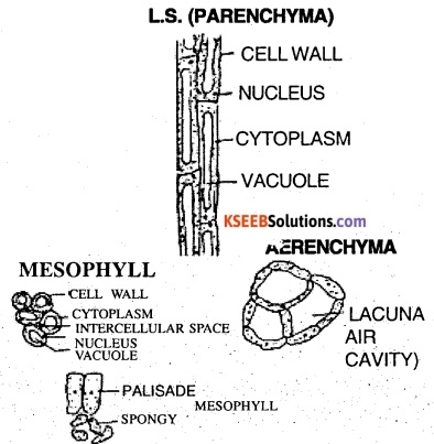
Functions:
- Parenchyma is basically a packing tissue and specialized tissues are embedded in it.
- Parenchyma act as a storage tissue and store variety of substances.
- In aquatic plants and petioles of canna and Musa they enclose large air cavities to help in buoyancy. Such parenchyma is called aerenchyma.
- With chloroplasts in the parenchyma it forms the chlorenchyma which helps in photosynthesis. The mesophyll of the leaf is made of this tissue.
- parenchyma cells when tightly packed and turgid provides mechanical support to herbaceous plants.
- cells can dedifferentiate and acquire the power of division.
The main type of parenchyma are
- Storage parenchyma: which helps in storage of starch, fats ergastic substances and water.
- Aerenchyma: which have large air cavities or intercellular spaces and provide buoyancy in aquatic plants.
- Chlorenchyma: contains chloroplasts carries on photosynthesis.
![]()
Question 2.
Describe briefly simple dead mechanical tissue. (Oct. 1985)
Answer:
Selerenchyma has its origin from the Gk word scleras meaning hard. The primary wall is made of cellulose and secondary wall is uniformly laid by a process called lignification. The cells are dead and enclose a central cavity called lumen. Certain places are unthickened and called pits. Selerenchyma is of 2 types fibres and selereids.
Fibres:
There are elongated, spindle shaped, with pointed needle like tips. They occur in groups have thick secondary walls and posses a narrow lumen. They are associated with roots, stems and leaves. Xylem fibres are found associated with xylem, phloem fibres with phloem, pericyclic fibres in the pericycle, cortical fibres in the cortex. They also surround the Vascular bundles of monocot stems. They help in providing mechanical support. They yield fibres of commerce (jute, hemp), provide protection to the vascular bundles.
Steroids or stone cells:
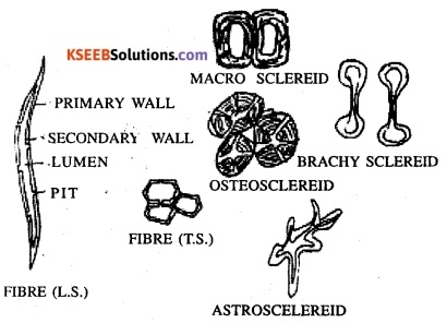
Vary in shape, thick and hard, with lignified walls showing concentric layering. Lumen is narrow and pits are also present. The grittiness of pulp of fruits like guava, pears is due to the occurrence of selereids, groups. Based on their shape they are grouped as Brachysclereids (stone cells), Macro Selereids (rod-shaped), Osteo Sclereids (bone shaped), and Astrosclereids (star-shaped). These provide mechanical support and rigidity to regions where they are present.
Question 3.
What are meristems? Classify meristems and give their functions. (Oct. 1994)
Answer:
A meristem is a group of undifferentiated cells, which are isodiametric and bring about rapid cell division. Meristems are classified based on position, origin and function. Based on position;
They are grouped into 3 types
- Apical Meristems: Found at the apices of roots and stems resulting in primary growth. They bring about an increase in the length of roots and stems.
- Intercalary Meristems: These are portions of apical meristems separated by permanent tissues. They occur at the base of internodes and leaf sheaths of monocots & grasses. They bring about elongation of the internode.
- Lateral Meristems: These occupy lateral positions and bring about an increase in diameter.
Based on Origin: They are grouped into 3 types
(a) Promeristem: These occupy the tip of stem and roots and represent a small area. By division, they give rise to primary meristems.
(b) Primary Meristems: These are derivatives of the premeristem and are found at the apices of shoot and root. They bring, about primary growth resulting in elongation of root & shoots.
(c) Secondary meristems; These are produced by dedifferentiation of permanent cells and arise later in the life of a plant. They occupy lateral positions and bring about secondary growth resulting in increase in diameter.
Based on Function: They are grouped into 3 types
- Protodprm: Single layered outer region which gives rise to the epidermis.
- Ground meristem: Many layered and forms the cortex and pith.
- Procambium: many-layered and forms the vascular cylinder.
Question 4.
With labelled diagrams, explain the kinds of xylem vessels (tracheae) based on their lignification. (March 2009, 2011)
Answer:
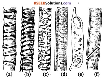
Xylem vessels tracheae are grouped into the followed types based on their lignification. The outer wall represents the primary wall and the inner lignified wall represents the secondary wall. Since the process of lignification uses all the cytoplasm the cells are dead and leaves behind a cavity called lumen. The xylem vessels tracheae show the following types based on lignification.
- Annular – where the secondary wall forms ring-like thickenings.
- Spiral – where the secondary wall forms spring-like thickenings.
- Reticulate – where the secondary wall forms a network like thickenings.
- Scalariform – where the secondary wall forms ladder-like thickenings.
- Pitted – where the secondary wall shows unthickened regions called pits.
- Bordered pitted – where the secondary wall shows bordered pits.
Question 5.
List any five characteristics of meristem. (March 2010)
Answer:
The characteristics of meristems are:
- All the cells are undifferentiated and isodiametric
- The cell wall is thin made of a primary wall of cellulose.
- The protoplast shows the presence of a prominent nucleus.
- The cytoplasm is filled with a number of small vacuoles.
- Cell organelles are absent and chloroplasts are in the proplastid stage.
- Cells are in an active stage of division.
Question 6.
Draw a neat labelled diagram of T.S. of a dorsiventral leaf and describe the different parts. (March 2008)
Answer:
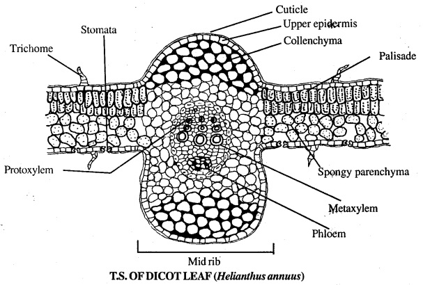
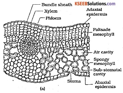
T.S. of dicot leaf (Eg: Helianthu The T.S. of dicot leaf shows three regions namely epidermis, mesophyll and the vascular bundles.
(i) Epidermis:
This layer is present on both upper and the lower surface of the leaf. It is composed of single layer of cells with deposition of cuticle. There are openings on the lower epidermis called stomata surrounded by the guard cells. The stomata open into an air space.
(ii) Mesophyll:
It is the tissue lying between the upper and the lower epidermis. It is further divided into two regions namely palisade parenchyma and spongy parenchyma. Palisade jarenchyma has about three layers of cells, which are tubular. These cells lie below the upper epidermis. There are air spaces here and there. Spongy parenchyma is composed of loosely arranged irregular cells. There are few layers of cells below the palisade parenchyma. Both palisade and spongy parenchyma cells contain abundant chloroplasts and help in photosynthesis
![]()
(iii) Vascular bundles :
These are conjoint, collateral and closed type. These are present in the midrib and vein regions. There is bundle sheath composed of parenchyma cells surrounding each vascular bundle. The cells are without chloroplasts. In the midrib region below the epidermis, there are collenchyma and parenchyma cell forming bundle sheath extensions.
Question 7.
Draw a neat labelled diagram of T.S. of Isobilateral leaf and explain the different parts. (July 2007) OR With a neat labelled diagram, explain the transverse section of monocot leaf.
Answer:
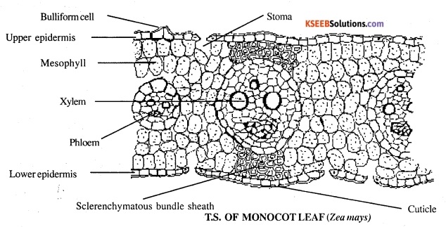
The T.S. of monocot leaf (Eg: Zee mays). The T.S. of monocot leaf shows three regions namely epidermis, mesophyll and vascular bundle.
(i) Epidermis:
There are two layers of epidermis namely upper and lower epidermis. Both the layers are composed of a single layer of parenchyma cells with deposition of the cuticle. There are openings in both the layers called stomata surrounded by guard cells. There are elongated cells on the upper epidermis called bulliform cells or motor cells, which help in rolling the leaf to check transpiration.
(ii) Mesophyll:
It is the tissue present between the two layers of the epidermis. It is composed of loosely arranged cells with lots of intercellular spaces. The cells have chloroplasts and are photosynthetic
(iii) Vascular bundles:
It is a conjoint, collateral and closed type with xylem facing upper epidermis and phloem facing the lower epidermis. There is no cambium between xylem and phloem. The vascular bundles are surrounded by bundle sheath of parenchyma cells.
Question 8.
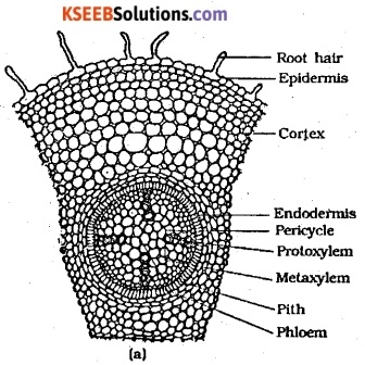
Draw a neat labelled diagram of Transverse Section of young ‘Dicot Root’ and explain different parts. (July 2006, 2010)
Answer:
The internal anatomy of root shows the following regions.
Epiblema or Piliferous Layer:
Single layered, compactly arranged cells, cuticle absent and provided with unicellular root hairs.
Cortex:
General Cortex is homogenously made of parenchyma. In old roots the epiblema dies off and the cells are suberised to form the exodermis. Innermost layer is the endodermis made of barrel-shaped cells with casparian thickenings. Endodermal cells opposite the protoxylem back the casparian thickenings and are called passage cells.
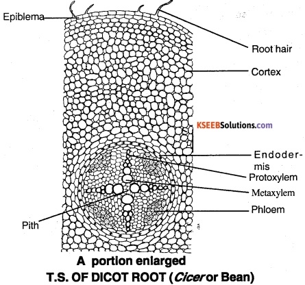
Stele:
Surrounded by a single-layered pericycle. Vascular bundles are limited in number i.e. diarch to search, usually tetrarch, radially arranged with exarch xylem and alternating patches of phloem and xylem. Conjunctive tissue lies between xylem and phloem which is parenchymatous. Pith is usually absent but if present small and parenchymatous.
![]()
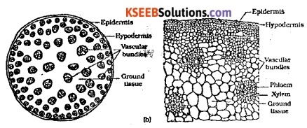
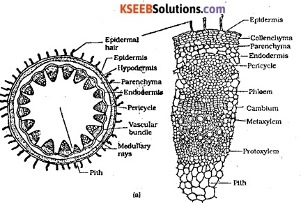
Question 9.
Differentiate between Monocot & dicot stem.
Answer:
| Monocot stem | Dicot stem |
| (1) Multicellular hairs absent | (1) Multicellular epidermal hair present |
| (2) Hypodermis sclerenchymatous | (2) Hypodermis Collenchymatous |
| (3) Vascular bundles numerous and scattered. (Atactostele) | (3) Vascular bundles limited, arranged as a broken ring (Eustele) |
| (4) Vascular bundles collateral, conjoint, closed, endarch | (4) Vascular bundles collateral, conjoint, open, endarch |
| (5) Endodermis, pericycle absent | (5) Endodermis, pericycle present |
| (6) Stem shows ground tissue | (6) Stem show cortex and pith |
| Monocot root | Dicot root |
| (1) Vascular bundles many, so-called polyarch | (1) Vascular bundles limited, most commonly tetrarch |
| (2) Casparian thickenings of endodermis ‘U’ shaped | (2) Casparian thickenings of endodermis are found on both radial and tangential walls |
| (3) Conjunctive tissue sclerenchymatous | (3) Conjunctive tissue parenchymatous |
| (4) Pith large, prominent & parenchymatous | (4) Pith absent, if present very small |